 ムービー
ムービー コントローラー


+ データを開く

- 基本情報
基本情報
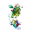
| 登録情報 | データベース: PDB / ID: 8gsy | ||||||
|---|---|---|---|---|---|---|---|
| タイトル | X-ray structure of Clostridium perfringens pili protein B N-terminal domain | ||||||
 要素 要素 | Peptidoglycan bound protein | ||||||
 キーワード キーワード | PROTEIN FIBRIL / Pili protein | ||||||
| 機能・相同性 | Collagen binding domain / Collagen binding domain / Fibrogen-binding domain 1 / Adhesion domain superfamily / collagen binding / cell adhesion / extracellular region / membrane / Peptidoglycan bound protein 機能・相同性情報 機能・相同性情報 | ||||||
| 生物種 |   Clostridium perfringens (ウェルシュ菌) Clostridium perfringens (ウェルシュ菌) | ||||||
| 手法 |  X線回折 / シンクロトロン / 分子置換 / 解像度: 1.55 Å X線回折 / シンクロトロン / 分子置換 / 解像度: 1.55 Å | ||||||
 データ登録者 データ登録者 | Kamitori, S. / Yamada, M. / Tamai, E. | ||||||
| 資金援助 |  日本, 1件 日本, 1件
| ||||||
 引用 引用 | ジャーナル: Febs Lett. / 年: 2023 タイトル: Structural and biochemical characterization of Clostridium perfringens pili protein B collagen-binding domains. 著者: Tamai, E. / Yamada, M. / Ishida, T. / Arimura, N. / Matsunami, R. / Sekiya, H. / Kamitori, S. | ||||||
| 履歴 |
|
- 構造の表示
構造の表示
| 構造ビューア | 分子: MolmilJmol/JSmol |
|---|
- ダウンロードとリンク
-ダウンロード
| PDBx/mmCIF形式 | 8gsy.cif.gz | 115.3 KB | 表示 | PDBx/mmCIF形式 |
|---|---|---|---|---|
| PDB形式 | pdb8gsy.ent.gz | 88 KB | 表示 | PDB形式 |
| PDBx/mmJSON形式 | 8gsy.json.gz | ツリー表示 | PDBx/mmJSON形式 | |
| その他 |  その他のダウンロード その他のダウンロード |
-検証レポート
| アーカイブディレクトリ | https://data.pdbj.org/pub/pdb/validation_reports/gs/8gsyftp://data.pdbj.org/pub/pdb/validation_reports/gs/8gsy | HTTPS FTP |
|---|
-関連構造データ
-リンク
 PDBj
PDBj- 集合体
集合体
| 登録構造単位 | 
| ||||||||
|---|---|---|---|---|---|---|---|---|---|
| 1 | 
| ||||||||
| 2 | 
| ||||||||
| 単位格子 |
|
-要素
| #1: タンパク質 | 分子量: 16368.459 Da / 分子数: 2 / 断片: N-terminal domain / 由来タイプ: 組換発現 由来: (組換発現) Clostridium perfringens (ウェルシュ菌)遺伝子: NCTC8081_03081 / 発現宿主: #2: 水 | ChemComp-HOH / |  分子量: 18.015 Da / 分子数: 104 / 由来タイプ: 天然 / 式: H2O 分子量: 18.015 Da / 分子数: 104 / 由来タイプ: 天然 / 式: H2O |
|---|
-実験情報
-実験
| 実験 | 手法: X線回折 / 使用した結晶の数: 1 |
|---|
- 試料調製
試料調製
| 結晶 | マシュー密度: 1.96 Å3/Da / 溶媒含有率: 37.14 % |
|---|---|
| 結晶化 | 温度: 293 K / 手法: 蒸気拡散法 詳細: 50 mM bis-tris pH 6.5, 50 mM ammonium sulfate, 30% v/v pentaerythritol ethoxylate (15/4 EO/OH) |
-データ収集
| 回折 | 平均測定温度: 100 K / Serial crystal experiment: N |
|---|---|
| 放射光源 | 由来: シンクロトロン / サイト: Photon Factory / ビームライン: BL-5A / 波長: 1 Å |
| 検出器 | タイプ: DECTRIS PILATUS3 6M / 検出器: PIXEL / 日付: 2022年3月22日 |
| 放射 | プロトコル: SINGLE WAVELENGTH / 単色(M)・ラウエ(L): M / 散乱光タイプ: x-ray |
| 放射波長 | 波長: 1 Å / 相対比: 1 |
| 反射 | 解像度: 1.55→48.48 Å / Num. obs: 39168 / % possible obs: 99.98 % / 冗長度: 9.4 % / CC1/2: 0.999 / Net I/σ(I): 23.23 |
| 反射 シェル | 解像度: 1.55→1.59 Å / Num. unique obs: 2809 / CC1/2: 0.789 |
- 解析
解析
| ソフトウェア |
| ||||||||||||||||||||||||||||||||||||||||||||||||||||||||||||||||||||||||||||||||||||||||||||||||||||||||||||||||||||||||||||||||||||||||||||||||||||||||||||||||||||||||||||||||||||||
|---|---|---|---|---|---|---|---|---|---|---|---|---|---|---|---|---|---|---|---|---|---|---|---|---|---|---|---|---|---|---|---|---|---|---|---|---|---|---|---|---|---|---|---|---|---|---|---|---|---|---|---|---|---|---|---|---|---|---|---|---|---|---|---|---|---|---|---|---|---|---|---|---|---|---|---|---|---|---|---|---|---|---|---|---|---|---|---|---|---|---|---|---|---|---|---|---|---|---|---|---|---|---|---|---|---|---|---|---|---|---|---|---|---|---|---|---|---|---|---|---|---|---|---|---|---|---|---|---|---|---|---|---|---|---|---|---|---|---|---|---|---|---|---|---|---|---|---|---|---|---|---|---|---|---|---|---|---|---|---|---|---|---|---|---|---|---|---|---|---|---|---|---|---|---|---|---|---|---|---|---|---|---|---|
| 精密化 | 構造決定の手法: 分子置換 開始モデル: 8GSX 解像度: 1.55→48.48 Å / Cor.coef. Fo:Fc: 0.968 / Cor.coef. Fo:Fc free: 0.947 / SU B: 4.51 / SU ML: 0.077 / 交差検証法: THROUGHOUT / ESU R: 0.096 / ESU R Free: 0.09 / 立体化学のターゲット値: MAXIMUM LIKELIHOOD / 詳細: HYDROGENS HAVE BEEN ADDED IN THE RIDING POSITIONS
| ||||||||||||||||||||||||||||||||||||||||||||||||||||||||||||||||||||||||||||||||||||||||||||||||||||||||||||||||||||||||||||||||||||||||||||||||||||||||||||||||||||||||||||||||||||||
| 溶媒の処理 | イオンプローブ半径: 0.8 Å / 減衰半径: 0.8 Å / VDWプローブ半径: 1.2 Å / 溶媒モデル: MASK | ||||||||||||||||||||||||||||||||||||||||||||||||||||||||||||||||||||||||||||||||||||||||||||||||||||||||||||||||||||||||||||||||||||||||||||||||||||||||||||||||||||||||||||||||||||||
| 原子変位パラメータ | Biso mean: 31.172 Å2
| ||||||||||||||||||||||||||||||||||||||||||||||||||||||||||||||||||||||||||||||||||||||||||||||||||||||||||||||||||||||||||||||||||||||||||||||||||||||||||||||||||||||||||||||||||||||
| 精密化ステップ | サイクル: 1 / 解像度: 1.55→48.48 Å
| ||||||||||||||||||||||||||||||||||||||||||||||||||||||||||||||||||||||||||||||||||||||||||||||||||||||||||||||||||||||||||||||||||||||||||||||||||||||||||||||||||||||||||||||||||||||
| 拘束条件 |
|