 Movie
Movie Controller


+ Open data

- Basic information
| Entry | Database: PDB / ID: 8gsc | |||||||||||||||||||||||||||||||||
|---|---|---|---|---|---|---|---|---|---|---|---|---|---|---|---|---|---|---|---|---|---|---|---|---|---|---|---|---|---|---|---|---|---|---|
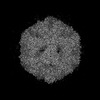
| Title | Echovirus3 A-particle in complex with 6D10 Fab | |||||||||||||||||||||||||||||||||
 Components Components |
| |||||||||||||||||||||||||||||||||
 Keywords Keywords | VIRUS/IMMUNE SYSTEM / E3 Full particle / Echovirus3 Full particle / antibody / 6D10 / VIRUS-IMMUNE SYSTEM complex | |||||||||||||||||||||||||||||||||
| Function / homology |  Function and homology information Function and homology informationsymbiont-mediated suppression of host cytoplasmic pattern recognition receptor signaling pathway via inhibition of RIG-I activity / picornain 2A / symbiont-mediated suppression of host mRNA export from nucleus / symbiont genome entry into host cell via pore formation in plasma membrane / picornain 3C / T=pseudo3 icosahedral viral capsid / host cell cytoplasmic vesicle membrane / viral capsid / host cell / nucleoside-triphosphate phosphatase ...symbiont-mediated suppression of host cytoplasmic pattern recognition receptor signaling pathway via inhibition of RIG-I activity / picornain 2A / symbiont-mediated suppression of host mRNA export from nucleus / symbiont genome entry into host cell via pore formation in plasma membrane / picornain 3C / T=pseudo3 icosahedral viral capsid / host cell cytoplasmic vesicle membrane / viral capsid / host cell / nucleoside-triphosphate phosphatase / channel activity / monoatomic ion transmembrane transport / DNA replication / RNA helicase activity / endocytosis involved in viral entry into host cell / symbiont-mediated activation of host autophagy / RNA-directed RNA polymerase / cysteine-type endopeptidase activity / viral RNA genome replication / RNA-directed RNA polymerase activity / DNA-templated transcription / symbiont entry into host cell / virion attachment to host cell / host cell nucleus / structural molecule activity / ATP hydrolysis activity / proteolysis / RNA binding / zinc ion binding / ATP binding / membrane Similarity search - Function | |||||||||||||||||||||||||||||||||
| Biological species |   Echovirus E3 Echovirus E3 | |||||||||||||||||||||||||||||||||
| Method | ELECTRON MICROSCOPY / single particle reconstruction / cryo EM / Resolution: 3.4 Å | |||||||||||||||||||||||||||||||||
 Authors Authors | Wang, X. / Fu, W. | |||||||||||||||||||||||||||||||||
| Funding support | 1items
| |||||||||||||||||||||||||||||||||
 Citation Citation | Journal: Viruses / Year: 2022 Title: Structural Basis for the Immunogenicity of the C-Terminus of VP1 of Echovirus 3 Revealed by the Binding of a Neutralizing Antibody. Authors: Shuai Qi / Wangjun Fu / Jinyan Fan / Li Zhang / Binyang Zheng / Kang Wang / Xiangxi Wang / Ling Zhu / Xinjian Li / Yuxia Zhang /  Abstract: Echovirus 3 (E3), a serotype of human enterovirus B (HEV-B), causes severe diseases in infants. Here, we determined the structures of E3 with a monoclonal antibody (MAb) 6D10 by cryo-EM to ...Echovirus 3 (E3), a serotype of human enterovirus B (HEV-B), causes severe diseases in infants. Here, we determined the structures of E3 with a monoclonal antibody (MAb) 6D10 by cryo-EM to comprehensively understand the specificities and the immunological characteristic of this serotype. The solved cryo-EM structures of the F-, A-, and E-particles of E3 bound with 6D10 revealed the structural features of the virus-antibody interface. Importantly, the structures of E-particles bound with 6D10 revealed for the first time the nature of the C-terminus of VP1 for HEV-Bs at the structural level. The highly immunogenic nature of this region in the E-particles provides new strategies for vaccine development for HEV-Bs. | |||||||||||||||||||||||||||||||||
| History |
|
- Structure visualization
| Structure viewer | Molecule: MolmilJmol/JSmol |
|---|
- Downloads & links
-Download
| PDBx/mmCIF format | 8gsc.cif.gz | 181.3 KB | Display | PDBx/mmCIF format |
|---|---|---|---|---|
| PDB format | pdb8gsc.ent.gz | 139.9 KB | Display | PDB format |
| PDBx/mmJSON format | 8gsc.json.gz | Tree view | PDBx/mmJSON format | |
| Others |  Other downloads Other downloads |
-Validation report
| Summary document | 8gsc_validation.pdf.gz | 1.6 MB | Display | wwPDB validaton report |
|---|---|---|---|---|
| Full document | 8gsc_full_validation.pdf.gz | 1.6 MB | Display | |
| Data in XML | 8gsc_validation.xml.gz | 39 KB | Display | |
| Data in CIF | 8gsc_validation.cif.gz | 57.1 KB | Display | |
| Arichive directory | https://data.pdbj.org/pub/pdb/validation_reports/gs/8gscftp://data.pdbj.org/pub/pdb/validation_reports/gs/8gsc | HTTPS FTP |
-Related structure data
| Related structure data | 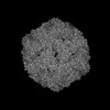 34231MC 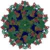 8gsdC 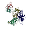 8gseC 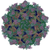 8gsfC M: map data used to model this data C: citing same article ( |
|---|---|
| Similar structure data |


-Links
 PDBj
PDBj


- Assembly
| Deposited unit | 
|
|---|---|
| 1 | x 60
|
| 2 |
|
| 3 | x 5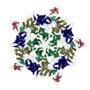
|
| 4 | x 6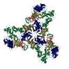
|
| 5 | 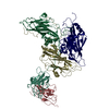
|
| Symmetry | Point symmetry: (Schoenflies symbol: I (icosahedral)) |
-Components
| #1: Protein | Mass: 26940.322 Da / Num. of mol.: 1 / Source method: isolated from a natural source / Source: (natural) Echovirus E3 / References: UniProt: A0A060BFD5 |
|---|---|
| #2: Protein | Mass: 27716.184 Da / Num. of mol.: 1 / Source method: isolated from a natural source / Source: (natural) Echovirus E3 / References: UniProt: A0A6M4MJE3 |
| #3: Protein | Mass: 25896.449 Da / Num. of mol.: 1 / Source method: isolated from a natural source / Source: (natural) Echovirus E3 / References: UniProt: A0A0K0LDT3 |
| #4: Protein | Mass: 12938.200 Da / Num. of mol.: 1 Source method: isolated from a genetically manipulated source Source: (gene. exp.) |
| #5: Antibody | Mass: 12652.158 Da / Num. of mol.: 1 Source method: isolated from a genetically manipulated source Source: (gene. exp.) |
| Has protein modification | Y |
-Experimental details
-Experiment
| Experiment | Method: ELECTRON MICROSCOPY |
|---|---|
| EM experiment | Aggregation state: PARTICLE / 3D reconstruction method: single particle reconstruction |
- Sample preparation
| Component |
| ||||||||||||||||||||||||
|---|---|---|---|---|---|---|---|---|---|---|---|---|---|---|---|---|---|---|---|---|---|---|---|---|---|
| Source (natural) |
| ||||||||||||||||||||||||
| Source (recombinant) | Organism: | ||||||||||||||||||||||||
| Details of virus | Empty: NO / Enveloped: YES / Isolate: OTHER / Type: VIRION | ||||||||||||||||||||||||
| Buffer solution | pH: 7.4 | ||||||||||||||||||||||||
| Specimen | Embedding applied: NO / Shadowing applied: NO / Staining applied: NO / Vitrification applied: YES | ||||||||||||||||||||||||
| Vitrification | Cryogen name: ETHANE |
- Electron microscopy imaging
| Experimental equipment |  Model: Titan Krios / Image courtesy: FEI Company |
|---|---|
| Microscopy | Model: FEI TITAN KRIOS |
| Electron gun | Electron source:  FIELD EMISSION GUN / Accelerating voltage: 300 kV / Illumination mode: FLOOD BEAM FIELD EMISSION GUN / Accelerating voltage: 300 kV / Illumination mode: FLOOD BEAM |
| Electron lens | Mode: DARK FIELD / Nominal defocus max: 2500 nm / Nominal defocus min: 1200 nm |
| Image recording | Electron dose: 30 e/Å2 / Film or detector model: GATAN K2 QUANTUM (4k x 4k) |
- Processing
| Software | Name: PHENIX / Version: 1.19.2_4158: / Classification: refinement | ||||||||||||||||||||||||
|---|---|---|---|---|---|---|---|---|---|---|---|---|---|---|---|---|---|---|---|---|---|---|---|---|---|
| EM software | Name: PHENIX / Category: model refinement | ||||||||||||||||||||||||
| CTF correction | Type: NONE | ||||||||||||||||||||||||
| 3D reconstruction | Resolution: 3.4 Å / Resolution method: FSC 0.143 CUT-OFF / Num. of particles: 55687 / Symmetry type: POINT | ||||||||||||||||||||||||
| Refine LS restraints |
|
