 Movie
Movie Controller

[English] 日本語
 Yorodumi
- PDB-8gs1: Crystal structure of AziU2-U3 complex from Streptomyces sahachiro... -
+ Open data

- Basic information
| Entry | Database: PDB / ID: 8gs1 | |||||||||
|---|---|---|---|---|---|---|---|---|---|---|
| Title | Crystal structure of AziU2-U3 complex from Streptomyces sahachiroi NRRL2485 | |||||||||
 Components Components |
| |||||||||
 Keywords Keywords | BIOSYNTHETIC PROTEIN / azinomycin B biosynthetic protein | |||||||||
| Function / homology | Butirosin biosynthesis protein H, N-terminal / Butirosin biosynthesis protein H, N-terminal / FORMIC ACID / LYSINE / Azi28 / Azi29 Function and homology information Function and homology information | |||||||||
| Biological species |  Streptomyces sahachiroi (bacteria) Streptomyces sahachiroi (bacteria) | |||||||||
| Method |  X-RAY DIFFRACTION / SYNCHROTRON / SAD / Resolution: 2.7 Å X-RAY DIFFRACTION / SYNCHROTRON / SAD / Resolution: 2.7 Å | |||||||||
 Authors Authors | Cheng, Y. / Li, P. / Liu, W. / Fang, P. | |||||||||
| Funding support |  China, 2items China, 2items
| |||||||||
 Citation Citation | Journal: J.Am.Chem.Soc. / Year: 2023 Title: Oxidase Heterotetramer Completes 1-Azabicyclo[3.1.0]hexane Formation with the Association of a Nonribosomal Peptide Synthetase. Authors: Cheng, Y. / Yi, X. / Zhang, Y. / He, Q. / Chen, D. / Cao, W. / Fang, P. / Liu, W. | |||||||||
| History |
|
- Structure visualization
| Structure viewer | Molecule: MolmilJmol/JSmol |
|---|
- Downloads & links
-Download
| PDBx/mmCIF format | 8gs1.cif.gz | 436.3 KB | Display | PDBx/mmCIF format |
|---|---|---|---|---|
| PDB format | pdb8gs1.ent.gz | 359.5 KB | Display | PDB format |
| PDBx/mmJSON format | 8gs1.json.gz | Tree view | PDBx/mmJSON format | |
| Others |  Other downloads Other downloads |
-Validation report
| Arichive directory | https://data.pdbj.org/pub/pdb/validation_reports/gs/8gs1ftp://data.pdbj.org/pub/pdb/validation_reports/gs/8gs1 | HTTPS FTP |
|---|
-Related structure data

-Links
 PDBj
PDBj- Assembly
| Deposited unit | 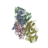
| ||||||||
|---|---|---|---|---|---|---|---|---|---|
| 1 | 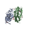
| ||||||||
| 2 | 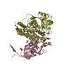
| ||||||||
| Unit cell |
| ||||||||
| Components on special symmetry positions |
|
-Components
| #1: Protein | Mass: 24087.084 Da / Num. of mol.: 2 Source method: isolated from a genetically manipulated source Source: (gene. exp.) Streptomyces sahachiroi (bacteria) / Gene: azi28 / Production host: #2: Protein | Mass: 38319.938 Da / Num. of mol.: 2 Source method: isolated from a genetically manipulated source Source: (gene. exp.) Streptomyces sahachiroi (bacteria) / Gene: azi29 / Production host: #3: Chemical | ChemComp-FMT /   Mass: 46.025 Da / Num. of mol.: 21 / Source method: obtained synthetically / Formula: CH2O2 / Feature type: SUBJECT OF INVESTIGATION Mass: 46.025 Da / Num. of mol.: 21 / Source method: obtained synthetically / Formula: CH2O2 / Feature type: SUBJECT OF INVESTIGATION#4: Chemical |   Type: L-peptide linking / Mass: 147.195 Da / Num. of mol.: 2 / Source method: isolated from a natural source / Formula: C6H15N2O2 Type: L-peptide linking / Mass: 147.195 Da / Num. of mol.: 2 / Source method: isolated from a natural source / Formula: C6H15N2O2#5: Water | ChemComp-HOH / |  Mass: 18.015 Da / Num. of mol.: 411 / Source method: isolated from a natural source / Formula: H2O Mass: 18.015 Da / Num. of mol.: 411 / Source method: isolated from a natural source / Formula: H2OHas ligand of interest | Y | |
|---|
-Experimental details
-Experiment
| Experiment | Method: X-RAY DIFFRACTION / Number of used crystals: 1 |
|---|
- Sample preparation
| Crystal | Density Matthews: 4.62 Å3/Da / Density % sol: 73.4 % |
|---|---|
| Crystal grow | Temperature: 291 K / Method: vapor diffusion, sitting drop / Details: Sodium formate, PEG 3350 |
-Data collection
| Diffraction | Mean temperature: 100 K / Serial crystal experiment: N | ||||||||||||||||||||||||||||||
|---|---|---|---|---|---|---|---|---|---|---|---|---|---|---|---|---|---|---|---|---|---|---|---|---|---|---|---|---|---|---|---|
| Diffraction source | Source: SYNCHROTRON / Site: SSRF / Beamline: BL18U1 / Wavelength: 0.97915 Å | ||||||||||||||||||||||||||||||
| Detector | Type: DECTRIS PILATUS 6M / Detector: PIXEL / Date: Jan 8, 2021 | ||||||||||||||||||||||||||||||
| Radiation | Protocol: SINGLE WAVELENGTH / Monochromatic (M) / Laue (L): M / Scattering type: x-ray | ||||||||||||||||||||||||||||||
| Radiation wavelength | Wavelength: 0.97915 Å / Relative weight: 1 | ||||||||||||||||||||||||||||||
| Reflection | Resolution: 2.7→50 Å / Num. obs: 122616 / % possible obs: 99.7 % / Redundancy: 19.8 % / Biso Wilson estimate: 50.21 Å2 / CC1/2: 0.995 / Rmerge(I) obs: 0.309 / Rpim(I) all: 0.07 / Rrim(I) all: 0.317 / Net I/σ(I): 11.1 | ||||||||||||||||||||||||||||||
| Reflection shell | Diffraction-ID: 1
|
- Processing
| Software |
| ||||||||||||||||||||||||||||||||||||||||||||||||||||||||||||||||||||||||
|---|---|---|---|---|---|---|---|---|---|---|---|---|---|---|---|---|---|---|---|---|---|---|---|---|---|---|---|---|---|---|---|---|---|---|---|---|---|---|---|---|---|---|---|---|---|---|---|---|---|---|---|---|---|---|---|---|---|---|---|---|---|---|---|---|---|---|---|---|---|---|---|---|---|
| Refinement | Method to determine structure: SAD / Resolution: 2.7→27.26 Å / SU ML: 0.37 / Cross valid method: THROUGHOUT / σ(F): 0.34 / Phase error: 24.6 / Stereochemistry target values: ML
| ||||||||||||||||||||||||||||||||||||||||||||||||||||||||||||||||||||||||
| Solvent computation | Shrinkage radii: 0.9 Å / VDW probe radii: 1.1 Å / Solvent model: FLAT BULK SOLVENT MODEL | ||||||||||||||||||||||||||||||||||||||||||||||||||||||||||||||||||||||||
| Displacement parameters | Biso max: 124.09 Å2 / Biso mean: 54.8501 Å2 / Biso min: 34.7 Å2 | ||||||||||||||||||||||||||||||||||||||||||||||||||||||||||||||||||||||||
| Refinement step | Cycle: final / Resolution: 2.7→27.26 Å
| ||||||||||||||||||||||||||||||||||||||||||||||||||||||||||||||||||||||||
| LS refinement shell | Refine-ID: X-RAY DIFFRACTION / Rfactor Rfree error: 0 / Total num. of bins used: 10 / % reflection obs: 100 %
| ||||||||||||||||||||||||||||||||||||||||||||||||||||||||||||||||||||||||
| Refinement TLS params. | Method: refined / Origin x: 47.1481 Å / Origin y: 136.3369 Å / Origin z: 85.0191 Å
| ||||||||||||||||||||||||||||||||||||||||||||||||||||||||||||||||||||||||
| Refinement TLS group |
|
