 Movie
Movie Controller


+ Open data

- Basic information
| Entry | Database: PDB / ID: 8gpm | ||||||
|---|---|---|---|---|---|---|---|
| Title | Acinetobacter baumannii carbonic anhydrase | ||||||
 Components Components | Carbonic anhydrase | ||||||
 Keywords Keywords | CARBOHYDRATE / Acinetobacter baumannii / carbonic anhydrase / trimer | ||||||
| Function / homology |  Function and homology information Function and homology information | ||||||
| Biological species |  Acinetobacter baumannii (bacteria) Acinetobacter baumannii (bacteria) | ||||||
| Method |  X-RAY DIFFRACTION / SYNCHROTRON / MOLECULAR REPLACEMENT / Resolution: 1.7 Å X-RAY DIFFRACTION / SYNCHROTRON / MOLECULAR REPLACEMENT / Resolution: 1.7 Å | ||||||
 Authors Authors | Wen, Y. / Jiao, M. | ||||||
| Funding support |  China, 1items China, 1items
| ||||||
 Citation Citation | Journal: Structure / Year: 2023 Title: Mechanistic and structural insights into the bifunctional enzyme PaaY from Acinetobacter baumannii. Authors: Jiao, M. / He, W. / Ouyang, Z. / Qin, Q. / Guo, Y. / Zhang, J. / Bai, Y. / Guo, X. / Yu, Q. / She, J. / Hwang, P.M. / Zheng, F. / Wen, Y. | ||||||
| History |
|
- Structure visualization
| Structure viewer | Molecule: MolmilJmol/JSmol |
|---|
- Downloads & links
-Download
| PDBx/mmCIF format | 8gpm.cif.gz | 96.3 KB | Display | PDBx/mmCIF format |
|---|---|---|---|---|
| PDB format | pdb8gpm.ent.gz | 72 KB | Display | PDB format |
| PDBx/mmJSON format | 8gpm.json.gz | Tree view | PDBx/mmJSON format | |
| Others |  Other downloads Other downloads |
-Validation report
| Summary document | 8gpm_validation.pdf.gz | 899.8 KB | Display | wwPDB validaton report |
|---|---|---|---|---|
| Full document | 8gpm_full_validation.pdf.gz | 901.1 KB | Display | |
| Data in XML | 8gpm_validation.xml.gz | 11.1 KB | Display | |
| Data in CIF | 8gpm_validation.cif.gz | 15.6 KB | Display | |
| Arichive directory | https://data.pdbj.org/pub/pdb/validation_reports/gp/8gpmftp://data.pdbj.org/pub/pdb/validation_reports/gp/8gpm | HTTPS FTP |
-Related structure data
| Related structure data | 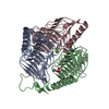 8gppC  1v3wS S: Starting model for refinement C: citing same article ( |
|---|---|
| Similar structure data |
-Links
 PDBj
PDBj


- Assembly
| Deposited unit | 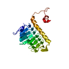
| ||||||||||||
|---|---|---|---|---|---|---|---|---|---|---|---|---|---|
| 1 | 
| ||||||||||||
| Unit cell |
| ||||||||||||
| Components on special symmetry positions |
|
-Components
| #1: Protein | Mass: 21896.869 Da / Num. of mol.: 1 Source method: isolated from a genetically manipulated source Source: (gene. exp.) Acinetobacter baumannii (bacteria) / Gene: paaY, ATCC19606_22550 / Production host: |
|---|---|
| #2: Chemical | ChemComp-GOL /   Mass: 92.094 Da / Num. of mol.: 1 / Source method: obtained synthetically / Formula: C3H8O3 Mass: 92.094 Da / Num. of mol.: 1 / Source method: obtained synthetically / Formula: C3H8O3 |
| #3: Chemical | ChemComp-ZN /   Mass: 65.409 Da / Num. of mol.: 1 / Source method: obtained synthetically / Formula: Zn / Feature type: SUBJECT OF INVESTIGATION Mass: 65.409 Da / Num. of mol.: 1 / Source method: obtained synthetically / Formula: Zn / Feature type: SUBJECT OF INVESTIGATION |
| #4: Chemical | ChemComp-CA /   Mass: 40.078 Da / Num. of mol.: 1 / Source method: obtained synthetically / Formula: Ca Mass: 40.078 Da / Num. of mol.: 1 / Source method: obtained synthetically / Formula: Ca |
| #5: Water | ChemComp-HOH /  Mass: 18.015 Da / Num. of mol.: 166 / Source method: isolated from a natural source / Formula: H2O Mass: 18.015 Da / Num. of mol.: 166 / Source method: isolated from a natural source / Formula: H2O |
| Has ligand of interest | Y |
| Has protein modification | N |
-Experimental details
-Experiment
| Experiment | Method: X-RAY DIFFRACTION / Number of used crystals: 1 |
|---|
- Sample preparation
| Crystal | Density Matthews: 2.44 Å3/Da / Density % sol: 49.67 % |
|---|---|
| Crystal grow | Temperature: 293 K / Method: vapor diffusion, sitting drop Details: 0.1 M bis-tris pH=6.5, 45% v/v polypropylene glycol P400 |
-Data collection
| Diffraction | Mean temperature: 100 K / Serial crystal experiment: N |
|---|---|
| Diffraction source | Source: SYNCHROTRON / Site: SSRF / Beamline: BL18U1 / Wavelength: 0.98 Å |
| Detector | Type: DECTRIS PILATUS 6M / Detector: PIXEL / Date: Nov 27, 2020 |
| Radiation | Protocol: SINGLE WAVELENGTH / Monochromatic (M) / Laue (L): M / Scattering type: x-ray |
| Radiation wavelength | Wavelength: 0.98 Å / Relative weight: 1 |
| Reflection | Resolution: 1.7→34.37 Å / Num. obs: 23637 / % possible obs: 99.92 % / Redundancy: 38.7 % / Biso Wilson estimate: 27.06 Å2 / CC1/2: 1 / Rmerge(I) obs: 0.101 / Net I/σ(I): 28.49 |
| Reflection shell | Resolution: 1.7→1.76 Å / Num. unique obs: 2337 / CC1/2: 0.832 |
- Processing
| Software |
| ||||||||||||||||||||||||||||||||||||||||||||||||||||||||||||||||||||||||||||||||||||||||||||||||||||||||||||||||||||||||||||||||||||||||||||||||||||||||||||||||||||||||
|---|---|---|---|---|---|---|---|---|---|---|---|---|---|---|---|---|---|---|---|---|---|---|---|---|---|---|---|---|---|---|---|---|---|---|---|---|---|---|---|---|---|---|---|---|---|---|---|---|---|---|---|---|---|---|---|---|---|---|---|---|---|---|---|---|---|---|---|---|---|---|---|---|---|---|---|---|---|---|---|---|---|---|---|---|---|---|---|---|---|---|---|---|---|---|---|---|---|---|---|---|---|---|---|---|---|---|---|---|---|---|---|---|---|---|---|---|---|---|---|---|---|---|---|---|---|---|---|---|---|---|---|---|---|---|---|---|---|---|---|---|---|---|---|---|---|---|---|---|---|---|---|---|---|---|---|---|---|---|---|---|---|---|---|---|---|---|---|---|---|
| Refinement | Method to determine structure: MOLECULAR REPLACEMENT Starting model: 1V3W Resolution: 1.7→34.37 Å / SU ML: 0.22 / Cross valid method: THROUGHOUT / σ(F): 1.37 / Phase error: 21.52 / Stereochemistry target values: ML
| ||||||||||||||||||||||||||||||||||||||||||||||||||||||||||||||||||||||||||||||||||||||||||||||||||||||||||||||||||||||||||||||||||||||||||||||||||||||||||||||||||||||||
| Solvent computation | Shrinkage radii: 0.9 Å / VDW probe radii: 1.11 Å / Solvent model: FLAT BULK SOLVENT MODEL | ||||||||||||||||||||||||||||||||||||||||||||||||||||||||||||||||||||||||||||||||||||||||||||||||||||||||||||||||||||||||||||||||||||||||||||||||||||||||||||||||||||||||
| Displacement parameters | Biso max: 109.55 Å2 / Biso mean: 31.3313 Å2 / Biso min: 17.7 Å2 | ||||||||||||||||||||||||||||||||||||||||||||||||||||||||||||||||||||||||||||||||||||||||||||||||||||||||||||||||||||||||||||||||||||||||||||||||||||||||||||||||||||||||
| Refinement step | Cycle: final / Resolution: 1.7→34.37 Å
| ||||||||||||||||||||||||||||||||||||||||||||||||||||||||||||||||||||||||||||||||||||||||||||||||||||||||||||||||||||||||||||||||||||||||||||||||||||||||||||||||||||||||
| LS refinement shell | Refine-ID: X-RAY DIFFRACTION / Rfactor Rfree error: 0 / Total num. of bins used: 27 / % reflection obs: 100 %
| ||||||||||||||||||||||||||||||||||||||||||||||||||||||||||||||||||||||||||||||||||||||||||||||||||||||||||||||||||||||||||||||||||||||||||||||||||||||||||||||||||||||||
| Refinement TLS params. | Method: refined / Origin x: 17.2172 Å / Origin y: -29.817 Å / Origin z: 11.1088 Å
| ||||||||||||||||||||||||||||||||||||||||||||||||||||||||||||||||||||||||||||||||||||||||||||||||||||||||||||||||||||||||||||||||||||||||||||||||||||||||||||||||||||||||
| Refinement TLS group |
|
