 Movie
Movie Controller

[English] 日本語
 Yorodumi
- PDB-8goa: Crystal Structure of Glycerol Dehydrogenase in the absence of NAD+ -
+ Open data

- Basic information
| Entry | Database: PDB / ID: 8goa | ||||||
|---|---|---|---|---|---|---|---|
| Title | Crystal Structure of Glycerol Dehydrogenase in the absence of NAD+ | ||||||
 Components Components | Glycerol dehydrogenase | ||||||
 Keywords Keywords | OXIDOREDUCTASE / Glycerol dehydrogenase / complex | ||||||
| Function / homology |  Function and homology information Function and homology information(R)-aminopropanol dehydrogenase / (R)-aminopropanol dehydrogenase activity / anaerobic glycerol catabolic process / methylglyoxal catabolic process / glycerol dehydrogenase / glycerol dehydrogenase (NAD+) activity / protein homotetramerization / protein-containing complex / zinc ion binding / identical protein binding / cytosol Similarity search - Function | ||||||
| Biological species |  | ||||||
| Method |  X-RAY DIFFRACTION / SYNCHROTRON / MOLECULAR REPLACEMENT / Resolution: 2.9 Å X-RAY DIFFRACTION / SYNCHROTRON / MOLECULAR REPLACEMENT / Resolution: 2.9 Å | ||||||
 Authors Authors | Park, T. / Hoang, H.N. / Kang, J.Y. / Park, J. / Mun, S.A. / Jin, M. / Yang, J. / Jung, C.-H. / Eom, S.H. | ||||||
| Funding support |  Korea, Republic Of, 1items Korea, Republic Of, 1items
| ||||||
 Citation Citation | Journal: Febs J. / Year: 2023 Title: Structural and functional insights into the flexible beta-hairpin of glycerol dehydrogenase. Authors: Park, T. / Hoang, H.N. / Kang, J.Y. / Park, J. / Mun, S.A. / Jin, M. / Yang, J. / Jung, C.H. / Eom, S.H. | ||||||
| History |
|
- Structure visualization
| Structure viewer | Molecule: MolmilJmol/JSmol |
|---|
- Downloads & links
-Download
| PDBx/mmCIF format | 8goa.cif.gz | 286.1 KB | Display | PDBx/mmCIF format |
|---|---|---|---|---|
| PDB format | pdb8goa.ent.gz | 231 KB | Display | PDB format |
| PDBx/mmJSON format | 8goa.json.gz | Tree view | PDBx/mmJSON format | |
| Others |  Other downloads Other downloads |
-Validation report
| Arichive directory | https://data.pdbj.org/pub/pdb/validation_reports/go/8goaftp://data.pdbj.org/pub/pdb/validation_reports/go/8goa | HTTPS FTP |
|---|
-Related structure data
| Related structure data | 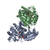 8gobC 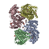 5zxlS S: Starting model for refinement C: citing same article ( |
|---|---|
| Similar structure data |
-Links
 PDBj
PDBj- Assembly
| Deposited unit | 
| ||||||||
|---|---|---|---|---|---|---|---|---|---|
| 1 | 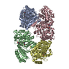
| ||||||||
| Unit cell |
|
-Components
| #1: Protein | Mass: 39817.152 Da / Num. of mol.: 4 Source method: isolated from a genetically manipulated source Source: (gene. exp.) #2: Chemical | ChemComp-ZN /   Mass: 65.409 Da / Num. of mol.: 4 / Source method: obtained synthetically / Formula: Zn / Feature type: SUBJECT OF INVESTIGATION Mass: 65.409 Da / Num. of mol.: 4 / Source method: obtained synthetically / Formula: Zn / Feature type: SUBJECT OF INVESTIGATION#3: Chemical | ChemComp-TRS /   Mass: 122.143 Da / Num. of mol.: 12 / Source method: obtained synthetically / Formula: C4H12NO3 / Feature type: SUBJECT OF INVESTIGATION / Comment: pH buffer*YM Mass: 122.143 Da / Num. of mol.: 12 / Source method: obtained synthetically / Formula: C4H12NO3 / Feature type: SUBJECT OF INVESTIGATION / Comment: pH buffer*YM#4: Water | ChemComp-HOH / |  Mass: 18.015 Da / Num. of mol.: 379 / Source method: isolated from a natural source / Formula: H2O Mass: 18.015 Da / Num. of mol.: 379 / Source method: isolated from a natural source / Formula: H2OHas ligand of interest | Y | |
|---|
-Experimental details
-Experiment
| Experiment | Method: X-RAY DIFFRACTION / Number of used crystals: 1 |
|---|
- Sample preparation
| Crystal | Density Matthews: 5.97 Å3/Da / Density % sol: 79.41 % |
|---|---|
| Crystal grow | Temperature: 293 K / Method: vapor diffusion, hanging drop Details: 100 mM sodium citrate (pH 5.6), 500 mM ammonium sulfate, and 1 M lithium sulfate |
-Data collection
| Diffraction | Mean temperature: 100 K / Serial crystal experiment: N |
|---|---|
| Diffraction source | Source: SYNCHROTRON / Site: PAL/PLS / Beamline: 11C / Wavelength: 0.9794 Å |
| Detector | Type: DECTRIS PILATUS3 6M / Detector: PIXEL / Date: Dec 14, 2019 |
| Radiation | Protocol: SINGLE WAVELENGTH / Monochromatic (M) / Laue (L): M / Scattering type: x-ray |
| Radiation wavelength | Wavelength: 0.9794 Å / Relative weight: 1 |
| Reflection | Resolution: 2.9→49.24 Å / Num. obs: 86081 / % possible obs: 99.6 % / Redundancy: 8.7 % / CC1/2: 0.99 / Net I/σ(I): 6.8 |
| Reflection shell | Resolution: 2.9→2.95 Å / Num. unique obs: 4204 / CC1/2: 0.322 |
- Processing
| Software |
| ||||||||||||||||||||||||
|---|---|---|---|---|---|---|---|---|---|---|---|---|---|---|---|---|---|---|---|---|---|---|---|---|---|
| Refinement | Method to determine structure: MOLECULAR REPLACEMENT Starting model: 5zxl Resolution: 2.9→49.24 Å / SU ML: 0.39 / Cross valid method: THROUGHOUT / σ(F): 1.33 / Phase error: 21.84 / Stereochemistry target values: ML
| ||||||||||||||||||||||||
| Solvent computation | Shrinkage radii: 0.9 Å / VDW probe radii: 1.1 Å / Solvent model: FLAT BULK SOLVENT MODEL | ||||||||||||||||||||||||
| Refinement step | Cycle: LAST / Resolution: 2.9→49.24 Å
| ||||||||||||||||||||||||
| Refine LS restraints |
| ||||||||||||||||||||||||
| LS refinement shell | Resolution: 2.9→2.93 Å
|
