[English] 日本語
Yorodumi
- PDB-8gl3: De novo design of monomeric helical bundles for pH-controlled mem... -

+
Open data


ID or keywords:

Loading...

-
Basic information

Entry
Database: PDB / ID: 8gl3
TitleDe novo design of monomeric helical bundles for pH-controlled membrane lysis
ComponentspRLB-519
KeywordsDE NOVO PROTEIN / protein design / pH-responsive / membrane lysis
Biological speciessynthetic construct (others)
MethodX-RAY DIFFRACTION / SYNCHROTRON / MOLECULAR REPLACEMENT / Resolution: 2.3 Å
AuthorsGoldbach, N. / Bera, A.K. / Baker, D. / Kang, A.
Funding support United States, 2items
OrganizationGrant numberCountry
National Science Foundation (NSF, United States) United States
Howard Hughes Medical Institute (HHMI) United States
CitationJournal: Protein Sci / Year: 2023
Title: De novo design of monomeric helical bundles for pH-controlled membrane lysis.
Authors: Nicolas Goldbach / Issa Benna / Basile I M Wicky / Jacob T Croft / Lauren Carter / Asim K Bera / Hannah Nguyen / Alex Kang / Banumathi Sankaran / Erin C Yang / Kelly K Lee / David Baker /
Abstract: Targeted intracellular delivery via receptor-mediated endocytosis requires the delivered cargo to escape the endosome to prevent lysosomal degradation. This can in principle be achieved by membrane ...Targeted intracellular delivery via receptor-mediated endocytosis requires the delivered cargo to escape the endosome to prevent lysosomal degradation. This can in principle be achieved by membrane lysis tightly restricted to endosomal membranes upon internalization to avoid general membrane insertion and lysis. Here, we describe the design of small monomeric proteins with buried histidine containing pH-responsive hydrogen bond networks and membrane permeating amphipathic helices. Of the 30 designs that were experimentally tested, all expressed in Escherichia coli, 13 were monomeric with the expected secondary structure, and 4 designs disrupted artificial liposomes in a pH-dependent manner. Mutational analysis showed that the buried histidine hydrogen bond networks mediate pH-responsiveness and control lysis of model membranes within a very narrow range of pH (6.0-5.5) with almost no lysis occurring at neutral pH. These tightly controlled lytic monomers could help mediate endosomal escape in designed targeted delivery platforms.
History
DepositionMar 20, 2023Deposition site: RCSB / Processing site: RCSB
Revision 1.0Sep 6, 2023Provider: repository / Type: Initial release
Revision 1.1Sep 20, 2023Group: Refinement description / Category: pdbx_initial_refinement_model / Item: _pdbx_initial_refinement_model.type
Revision 1.2Nov 8, 2023Group: Database references / Category: citation / Item: _citation.journal_volume

-
Structure visualization

Structure viewerMolecule:
MolmilJmol/JSmol

Downloads & links

-
Assembly

Deposited unit
A: pRLB-519


Theoretical massNumber of molelcules
Total (without water)27,5991
Polymers27,5991
Non-polymers00
Water55831
1


  • Idetical with deposited unit
  • defined by author
  • Evidence: SAXS
TypeNameSymmetry operationNumber
identity operation1_555x,y,z1
Unit cell
Length a, b, c (Å)32.120, 44.703, 182.579
Angle α, β, γ (deg.)90.000, 90.430, 90.000
Int Tables number5
Space group name H-MI121
Space group name HallC2y(x,y,-x+z)
Symmetry operation#1: x,y,z
#2: -x,y,-z
#3: x+1/2,y+1/2,z+1/2
#4: -x+1/2,y+1/2,-z+1/2

-
Components

#1: Protein pRLB-519


Mass: 27599.225 Da / Num. of mol.: 1
Source method: isolated from a genetically manipulated source
Source: (gene. exp.) synthetic construct (others) / Production host: Escherichia coli (E. coli)
#2: Water ChemComp-HOH / water


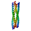
Mass: 18.015 Da / Num. of mol.: 31 / Source method: isolated from a natural source / Formula: H2O

-
Experimental details

-
Experiment

ExperimentMethod: X-RAY DIFFRACTION / Number of used crystals: 1

-
Sample preparation

CrystalDensity Matthews: 2.4 Å3/Da / Density % sol: 48.2 %
Crystal growTemperature: 293 K / Method: vapor diffusion, sitting drop / pH: 8 / Details: 0.2 M Sodium fluoride and 20% (w/v) PEG 3350

-
Data collection

DiffractionMean temperature: 100 K / Serial crystal experiment: N
Diffraction sourceSource: SYNCHROTRON / Site: ALS / Beamline: 8.2.2 / Wavelength: 0.99998 Å
DetectorType: DECTRIS PILATUS 6M / Detector: PIXEL / Date: Nov 4, 2022
RadiationProtocol: SINGLE WAVELENGTH / Monochromatic (M) / Laue (L): M / Scattering type: x-ray
Radiation wavelengthWavelength: 0.99998 Å / Relative weight: 1
ReflectionResolution: 2.3→45.64 Å / Num. obs: 11502 / % possible obs: 98.36 % / Redundancy: 3.3 % / Biso Wilson estimate: 23.78 Å2 / CC1/2: 0.998 / Rmerge(I) obs: 0.082 / Rpim(I) all: 0.052 / Net I/σ(I): 10.5
Reflection shellResolution: 2.3→2.53 Å / Redundancy: 3.4 % / Rmerge(I) obs: 0.144 / Mean I/σ(I) obs: 7.39 / Num. unique obs: 2812 / CC1/2: 0.976 / Rpim(I) all: 0.091 / % possible all: 98.13

-
Processing

Software
NameVersionClassification
PHENIX1.20.1_4487refinement
DIALSdata reduction
DIALSdata scaling
PHASERphasing
RefinementMethod to determine structure: MOLECULAR REPLACEMENT / Resolution: 2.3→45.64 Å / SU ML: 0.2172 / Cross valid method: FREE R-VALUE / σ(F): 1.42 / Phase error: 29.0929
Stereochemistry target values: GeoStd + Monomer Library + CDL v1.2
RfactorNum. reflection% reflection
Rfree0.2836 622 5.41 %
Rwork0.238 10880 -
obs0.2404 11502 98.37 %
Solvent computationShrinkage radii: 0.9 Å / VDW probe radii: 1.1 Å / Solvent model: FLAT BULK SOLVENT MODEL
Displacement parametersBiso mean: 46.04 Å2
Refinement stepCycle: LAST / Resolution: 2.3→45.64 Å
ProteinNucleic acidLigandSolventTotal
Num. atoms1600 0 0 31 1631
Refine LS restraints
Refine-IDTypeDev idealNumber
X-RAY DIFFRACTIONf_bond_d0.00161599
X-RAY DIFFRACTIONf_angle_d0.33682121
X-RAY DIFFRACTIONf_chiral_restr0.0264238
X-RAY DIFFRACTIONf_plane_restr0.0031285
X-RAY DIFFRACTIONf_dihedral_angle_d14.1537684
LS refinement shell
Resolution (Å)Rfactor RfreeNum. reflection RfreeRfactor RworkNum. reflection RworkRefine-ID% reflection obs (%)
2.3-2.530.30891550.23782658X-RAY DIFFRACTION98.22
2.53-2.90.26351670.24322676X-RAY DIFFRACTION97.2
2.9-3.650.28581420.22572768X-RAY DIFFRACTION99.76
3.65-45.640.28341580.24532778X-RAY DIFFRACTION98.29
Refinement TLS params.Method: refined / Origin x: 8.879 Å / Origin y: -7.312 Å / Origin z: 36.152 Å
111213212223313233
T0.130626554671 Å2-0.00505251078471 Å20.0213812019374 Å2-0.175752578267 Å2-0.0606928837649 Å2--0.183556758209 Å2
L2.45387639114 °2-0.491229070996 °2-0.831135296045 °2-2.200495235 °2-1.34783690293 °2--2.72451328743 °2
S0.0750647158982 Å °0.457226911539 Å °-0.0930397423271 Å °-0.176446076106 Å °0.0472748410748 Å °-0.0303733315338 Å °0.0857918009659 Å °-0.593079074475 Å °-0.0401827860087 Å °
Refinement TLS groupSelection details: ( CHAIN A AND RESID 6:221 )

+
About Yorodumi

-
News

-
Feb 9, 2022. New format data for meta-information of EMDB entries

New format data for meta-information of EMDB entries

  • Version 3 of the EMDB header file is now the official format.
  • The previous official version 1.9 will be removed from the archive.

Related info.:EMDB header

External links:wwPDB to switch to version 3 of the EMDB data model

-
Aug 12, 2020. Covid-19 info

Covid-19 info

URL: https://pdbjlvh1.pdbj.org/emnavi/covid19.php

New page: Covid-19 featured information page in EM Navigator.

Related info.:Covid-19 info / Mar 5, 2020. Novel coronavirus structure data

+
Mar 5, 2020. Novel coronavirus structure data

Novel coronavirus structure data

Related info.:Yorodumi Speices / Aug 12, 2020. Covid-19 info

External links:COVID-19 featured content - PDBj / Molecule of the Month (242):Coronavirus Proteases

+
Jan 31, 2019. EMDB accession codes are about to change! (news from PDBe EMDB page)

EMDB accession codes are about to change! (news from PDBe EMDB page)

  • The allocation of 4 digits for EMDB accession codes will soon come to an end. Whilst these codes will remain in use, new EMDB accession codes will include an additional digit and will expand incrementally as the available range of codes is exhausted. The current 4-digit format prefixed with “EMD-” (i.e. EMD-XXXX) will advance to a 5-digit format (i.e. EMD-XXXXX), and so on. It is currently estimated that the 4-digit codes will be depleted around Spring 2019, at which point the 5-digit format will come into force.
  • The EM Navigator/Yorodumi systems omit the EMD- prefix.

Related info.:Q: What is EMD? / ID/Accession-code notation in Yorodumi/EM Navigator

External links:EMDB Accession Codes are Changing Soon! / Contact to PDBj

+
Jul 12, 2017. Major update of PDB

Major update of PDB

  • wwPDB released updated PDB data conforming to the new PDBx/mmCIF dictionary.
  • This is a major update changing the version number from 4 to 5, and with Remediation, in which all the entries are updated.
  • In this update, many items about electron microscopy experimental information are reorganized (e.g. em_software).
  • Now, EM Navigator and Yorodumi are based on the updated data.

External links:wwPDB Remediation / Enriched Model Files Conforming to OneDep Data Standards Now Available in the PDB FTP Archive

-
Yorodumi

Thousand views of thousand structures

  • Yorodumi is a browser for structure data from EMDB, PDB, SASBDB, etc.
  • This page is also the successor to EM Navigator detail page, and also detail information page/front-end page for Omokage search.
  • The word "yorodu" (or yorozu) is an old Japanese word meaning "ten thousand". "mi" (miru) is to see.

Related info.:EMDB / PDB / SASBDB / Comparison of 3 databanks / Yorodumi Search / Aug 31, 2016. New EM Navigator & Yorodumi / Yorodumi Papers / Jmol/JSmol / Function and homology information / Changes in new EM Navigator and Yorodumi

Read more