+ Open data
- Basic information
| Entry | Database: PDB / ID: 8gkg | ||||||
|---|---|---|---|---|---|---|---|
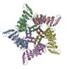
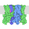
| Title | Human TRPV3 pentamer structure | ||||||
 Components Components | Transient receptor potential cation channel subfamily V member 3 | ||||||
 Keywords Keywords | MEMBRANE PROTEIN / TRP channel / ion channel / pore dilation | ||||||
| Function / homology |  Function and homology information Function and homology informationnegative regulation of hair cycle / osmosensory signaling pathway / response to temperature stimulus / TRP channels / positive regulation of calcium ion import / sodium channel activity / calcium ion import across plasma membrane / actin filament organization / calcium ion transmembrane transport / calcium channel activity ...negative regulation of hair cycle / osmosensory signaling pathway / response to temperature stimulus / TRP channels / positive regulation of calcium ion import / sodium channel activity / calcium ion import across plasma membrane / actin filament organization / calcium ion transmembrane transport / calcium channel activity / lysosome / receptor complex / cilium / metal ion binding / identical protein binding / plasma membrane / cytoplasm Similarity search - Function | ||||||
| Biological species |  Homo sapiens (human) Homo sapiens (human) | ||||||
| Method | ELECTRON MICROSCOPY / single particle reconstruction / cryo EM / Resolution: 4.38 Å | ||||||
 Authors Authors | Lansky, S. / Betancourt, J.M. / Scheuring, S. | ||||||
| Funding support |  United States, 1items United States, 1items
| ||||||
 Citation Citation |  Journal: Nature / Year: 2023 Journal: Nature / Year: 2023Title: A pentameric TRPV3 channel with a dilated pore. Authors: Shifra Lansky / John Michael Betancourt / Jingying Zhang / Yining Jiang / Elizabeth D Kim / Navid Paknejad / Crina M Nimigean / Peng Yuan / Simon Scheuring /  Abstract: Transient receptor potential (TRP) channels are a large, eukaryotic ion channel superfamily that control diverse physiological functions, and therefore are attractive drug targets. More than 210 ...Transient receptor potential (TRP) channels are a large, eukaryotic ion channel superfamily that control diverse physiological functions, and therefore are attractive drug targets. More than 210 structures from more than 20 different TRP channels have been determined, and all are tetramers. Despite this wealth of structures, many aspects concerning TRPV channels remain poorly understood, including the pore-dilation phenomenon, whereby prolonged activation leads to increased conductance, permeability to large ions and loss of rectification. Here, we used high-speed atomic force microscopy (HS-AFM) to analyse membrane-embedded TRPV3 at the single-molecule level and discovered a pentameric state. HS-AFM dynamic imaging revealed transience and reversibility of the pentamer in dynamic equilibrium with the canonical tetramer through membrane diffusive protomer exchange. The pentamer population increased upon diphenylboronic anhydride (DPBA) addition, an agonist that has been shown to induce TRPV3 pore dilation. On the basis of these findings, we designed a protein production and data analysis pipeline that resulted in a cryogenic-electron microscopy structure of the TRPV3 pentamer, showing an enlarged pore compared to the tetramer. The slow kinetics to enter and exit the pentameric state, the increased pentamer formation upon DPBA addition and the enlarged pore indicate that the pentamer represents the structural correlate of pore dilation. We thus show membrane diffusive protomer exchange as an additional mechanism for structural changes and conformational variability. Overall, we provide structural evidence for a non-canonical pentameric TRP-channel assembly, laying the foundation for new directions in TRP channel research. | ||||||
| History |
|
- Structure visualization
| Structure viewer | Molecule:  Molmil Molmil Jmol/JSmol Jmol/JSmol |
|---|
- Downloads & links
- Download
| PDBx/mmCIF format |  8gkg.cif.gz 8gkg.cif.gz | 431.6 KB | Display |  PDBx/mmCIF format PDBx/mmCIF format |
|---|---|---|---|---|
| PDB format |  pdb8gkg.ent.gz pdb8gkg.ent.gz | 323.1 KB | Display |  PDB format PDB format |
| PDBx/mmJSON format |  8gkg.json.gz 8gkg.json.gz | Tree view |  PDBx/mmJSON format PDBx/mmJSON format | |
| Others |  Other downloads Other downloads |
-Validation report
| Arichive directory |  https://data.pdbj.org/pub/pdb/validation_reports/gk/8gkg https://data.pdbj.org/pub/pdb/validation_reports/gk/8gkg ftp://data.pdbj.org/pub/pdb/validation_reports/gk/8gkg ftp://data.pdbj.org/pub/pdb/validation_reports/gk/8gkg | HTTPS FTP |
|---|
-Related structure data
| Related structure data |  40183MC  8gkaC C: citing same article ( M: map data used to model this data |
|---|---|
| Similar structure data | Similarity search - Function & homology  F&H Search F&H Search |
- Links
Links
- Assembly
| Deposited unit | 
|
|---|---|
| 1 |
|
- Components
| #1: Protein | Mass: 90742.812 Da / Num. of mol.: 5 Source method: isolated from a genetically manipulated source Source: (gene. exp.)  Homo sapiens (human) / Gene: TRPV3 / Cell line (production host): HEK293S GnTI- / Production host: Homo sapiens (human) / Gene: TRPV3 / Cell line (production host): HEK293S GnTI- / Production host:  Homo sapiens (human) / References: UniProt: Q8NET8 Homo sapiens (human) / References: UniProt: Q8NET8 |
|---|
-Experimental details
-Experiment
| Experiment | Method: ELECTRON MICROSCOPY |
|---|---|
| EM experiment | Aggregation state: PARTICLE / 3D reconstruction method: single particle reconstruction |
- Sample preparation
| Component | Name: TRPV3 ion channel, pentameric oligomerization assembly Type: COMPLEX / Entity ID: all / Source: RECOMBINANT | |||||||||||||||||||||||||
|---|---|---|---|---|---|---|---|---|---|---|---|---|---|---|---|---|---|---|---|---|---|---|---|---|---|---|
| Source (natural) | Organism:  Homo sapiens (human) Homo sapiens (human) | |||||||||||||||||||||||||
| Source (recombinant) | Organism:  Homo sapiens (human) / Cell: HEK293S GnTI- Homo sapiens (human) / Cell: HEK293S GnTI- | |||||||||||||||||||||||||
| Buffer solution | pH: 8 Details: 20 mM Tris pH 8, 150 mM NaCl, 0.01% GDN, 1 mM beta-mercaptoethanol | |||||||||||||||||||||||||
| Buffer component |
| |||||||||||||||||||||||||
| Specimen | Conc.: 3 mg/ml / Embedding applied: NO / Shadowing applied: NO / Staining applied: NO / Vitrification applied: YES | |||||||||||||||||||||||||
| Specimen support | Grid material: COPPER / Grid mesh size: 400 divisions/in. / Grid type: Quantifoil R1.2/1.3 | |||||||||||||||||||||||||
| Vitrification | Instrument: FEI VITROBOT MARK IV / Cryogen name: ETHANE / Humidity: 100 % / Chamber temperature: 277 K / Details: 3 ul sample blot 2 sec force 3 wait 20 sec |
- Electron microscopy imaging
| Experimental equipment |  Model: Titan Krios / Image courtesy: FEI Company |
|---|---|
| Microscopy | Model: FEI TITAN KRIOS |
| Electron gun | Electron source:  FIELD EMISSION GUN / Accelerating voltage: 300 kV / Illumination mode: FLOOD BEAM FIELD EMISSION GUN / Accelerating voltage: 300 kV / Illumination mode: FLOOD BEAM |
| Electron lens | Mode: BRIGHT FIELD / Nominal magnification: 64000 X / Nominal defocus max: 2500 nm / Nominal defocus min: 800 nm / Cs: 0.001 mm |
| Specimen holder | Cryogen: NITROGEN |
| Image recording | Electron dose: 53.27 e/Å2 / Film or detector model: GATAN K3 (6k x 4k) / Num. of grids imaged: 2 / Num. of real images: 10125 |
- Processing
| Software | Name: PHENIX / Version: 1.20.1_4487: / Classification: refinement | |||||||||||||||||||||||||||
|---|---|---|---|---|---|---|---|---|---|---|---|---|---|---|---|---|---|---|---|---|---|---|---|---|---|---|---|---|
| EM software |
| |||||||||||||||||||||||||||
| CTF correction | Type: PHASE FLIPPING AND AMPLITUDE CORRECTION | |||||||||||||||||||||||||||
| Particle selection | Num. of particles selected: 3668517 | |||||||||||||||||||||||||||
| Symmetry | Point symmetry: C5 (5 fold cyclic) | |||||||||||||||||||||||||||
| 3D reconstruction | Resolution: 4.38 Å / Resolution method: FSC 0.143 CUT-OFF / Num. of particles: 30186 / Symmetry type: POINT | |||||||||||||||||||||||||||
| Atomic model building | Protocol: FLEXIBLE FIT | |||||||||||||||||||||||||||
| Atomic model building | PDB-ID: 6UW4 Accession code: 6uw4 / Source name: PDB / Type: experimental model | |||||||||||||||||||||||||||
| Refine LS restraints |
|
 Movie
Movie Controller


 PDBj
PDBj


