 Movie
Movie Controller

[English] 日本語
 Yorodumi
- PDB-8gjc: X-ray crystallographic structure of a beta-hairpin peptide derive... -
+ Open data

- Basic information
| Entry | Database: PDB / ID: 8gjc | ||||||
|---|---|---|---|---|---|---|---|
| Title | X-ray crystallographic structure of a beta-hairpin peptide derived from Abeta 17-35. (ORN)LVFFAED(ORN)GAI(N-Me-Ile)GLM | ||||||
 Components Components | beta-hairpin peptide derived from Abeta 17-35 | ||||||
 Keywords Keywords | DE NOVO PROTEIN / oligomer / tetramer / amyloid / Alzheimer's disease | ||||||
| Function / homology | METHIONINE / DI(HYDROXYETHYL)ETHER / trifluoroacetic acid Function and homology information Function and homology information | ||||||
| Biological species | synthetic construct (others) | ||||||
| Method |  X-RAY DIFFRACTION / SYNCHROTRON / MOLECULAR REPLACEMENT / Resolution: 1.431 Å X-RAY DIFFRACTION / SYNCHROTRON / MOLECULAR REPLACEMENT / Resolution: 1.431 Å | ||||||
 Authors Authors | Kreutzer, A.G. / Ruttenberg, S.M. / Nowick, J.S. | ||||||
| Funding support |  United States, 1items United States, 1items
| ||||||
 Citation Citation | Journal: Biochemistry / Year: 2024 Title: beta-Hairpin Alignment Alters Oligomer Formation in A beta-Derived Peptides. Authors: Ruttenberg, S.M. / Kreutzer, A.G. / Truex, N.L. / Nowick, J.S. | ||||||
| History |
|
- Structure visualization
| Structure viewer | Molecule: MolmilJmol/JSmol |
|---|
- Downloads & links
-Download
| PDBx/mmCIF format | 8gjc.cif.gz | 25.3 KB | Display | PDBx/mmCIF format |
|---|---|---|---|---|
| PDB format | pdb8gjc.ent.gz | 17.1 KB | Display | PDB format |
| PDBx/mmJSON format | 8gjc.json.gz | Tree view | PDBx/mmJSON format | |
| Others |  Other downloads Other downloads |
-Validation report
| Arichive directory | https://data.pdbj.org/pub/pdb/validation_reports/gj/8gjcftp://data.pdbj.org/pub/pdb/validation_reports/gj/8gjc | HTTPS FTP |
|---|
-Related structure data
| Related structure data |  8gjdC 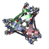 5w4hS S: Starting model for refinement C: citing same article ( |
|---|---|
| Similar structure data |
-Links
 PDBj
PDBj

- Assembly
| Deposited unit | 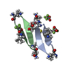
| ||||||||
|---|---|---|---|---|---|---|---|---|---|
| 1 | 
| ||||||||
| Unit cell |
| ||||||||
| Components on special symmetry positions |
|
-Components
| #1: Protein/peptide | Mass: 1606.903 Da / Num. of mol.: 2 / Source method: obtained synthetically / Source: (synth.) synthetic construct (others) #2: Chemical |   Type: L-peptide linking / Mass: 149.211 Da / Num. of mol.: 2 / Source method: obtained synthetically / Formula: C5H11NO2S Type: L-peptide linking / Mass: 149.211 Da / Num. of mol.: 2 / Source method: obtained synthetically / Formula: C5H11NO2S#3: Chemical |   Mass: 114.023 Da / Num. of mol.: 3 / Source method: obtained synthetically / Formula: C2HF3O2 Mass: 114.023 Da / Num. of mol.: 3 / Source method: obtained synthetically / Formula: C2HF3O2#4: Chemical | ChemComp-PEG / |   Mass: 106.120 Da / Num. of mol.: 1 / Source method: obtained synthetically / Formula: C4H10O3 Mass: 106.120 Da / Num. of mol.: 1 / Source method: obtained synthetically / Formula: C4H10O3#5: Water | ChemComp-HOH / |  Mass: 18.015 Da / Num. of mol.: 31 / Source method: isolated from a natural source / Formula: H2O Mass: 18.015 Da / Num. of mol.: 31 / Source method: isolated from a natural source / Formula: H2OHas ligand of interest | N | |
|---|
-Experimental details
-Experiment
| Experiment | Method: X-RAY DIFFRACTION / Number of used crystals: 1 |
|---|
- Sample preparation
| Crystal | Density Matthews: 2.1 Å3/Da / Density % sol: 41.34 % |
|---|---|
| Crystal grow | Temperature: 298.15 K / Method: vapor diffusion, hanging drop Details: Molecular Dynamics Morpheus solution containing 0.12M ethylene glycols mix, 0.1M Buffer System 3 pH 8.5, and 37.5% v/v precipitant mix 4. Molecular Dynamics ethylene glycols mix consists of ...Details: Molecular Dynamics Morpheus solution containing 0.12M ethylene glycols mix, 0.1M Buffer System 3 pH 8.5, and 37.5% v/v precipitant mix 4. Molecular Dynamics ethylene glycols mix consists of 0.3 M diethylene glycol, 0.3 M triethylene glycol, 0.3 M tetraethylene glycol, 0.3 M penta(ethylene glycol). Molecular Dynamics Buffer System 3 consists of 1 M BICINE and 1 M Trisma Base. Molecular Dynamics precipitant mix 4 consists of 25 % w/v hexylene glycol, 25 % w/v poly(ethylene glycol) 1000, and 25 % w/v poly(ethylene glycol) 3350. |
-Data collection
| Diffraction | Mean temperature: 103.15 K / Serial crystal experiment: N |
|---|---|
| Diffraction source | Source: SYNCHROTRON / Site: ALS / Beamline: 5.0.2 / Wavelength: 0.99999 Å |
| Detector | Type: DECTRIS PILATUS3 6M / Detector: PIXEL / Date: Apr 21, 2022 |
| Radiation | Protocol: SINGLE WAVELENGTH / Monochromatic (M) / Laue (L): M / Scattering type: x-ray |
| Radiation wavelength | Wavelength: 0.99999 Å / Relative weight: 1 |
| Reflection | Resolution: 1.431→23.01 Å / Num. obs: 9354 / % possible obs: 97.02 % / Redundancy: 16 % / CC1/2: 0.999 / Rmerge(I) obs: 0.09013 / Net I/σ(I): 24.44 |
| Reflection shell | Resolution: 1.431→1.482 Å / Rmerge(I) obs: 1.775 / Num. unique obs: 384 / CC1/2: 0.25 |
- Processing
| Software |
| ||||||||||||||||||||||||||||||||||||||||||||||||||||||||
|---|---|---|---|---|---|---|---|---|---|---|---|---|---|---|---|---|---|---|---|---|---|---|---|---|---|---|---|---|---|---|---|---|---|---|---|---|---|---|---|---|---|---|---|---|---|---|---|---|---|---|---|---|---|---|---|---|---|
| Refinement | Method to determine structure: MOLECULAR REPLACEMENT Starting model: 5W4H Resolution: 1.431→23.01 Å / Cross valid method: FREE R-VALUE / σ(F): 7.36 / Phase error: 20.89 / Stereochemistry target values: TWIN_LSQ_F
| ||||||||||||||||||||||||||||||||||||||||||||||||||||||||
| Solvent computation | Shrinkage radii: 0.9 Å / VDW probe radii: 1.1 Å / Solvent model: FLAT BULK SOLVENT MODEL | ||||||||||||||||||||||||||||||||||||||||||||||||||||||||
| Refinement step | Cycle: LAST / Resolution: 1.431→23.01 Å
| ||||||||||||||||||||||||||||||||||||||||||||||||||||||||
| Refine LS restraints |
| ||||||||||||||||||||||||||||||||||||||||||||||||||||||||
| LS refinement shell |
|
