 Movie
Movie Controller

[English] 日本語
 Yorodumi
- PDB-8gif: Crystal structure of a designed single-component Plasmodium falci... -
+ Open data

- Basic information
| Entry | Database: PDB / ID: 8gif | ||||||
|---|---|---|---|---|---|---|---|
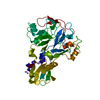
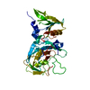
| Title | Crystal structure of a designed single-component Plasmodium falciparum AMA1-RON2L insertion fusion immunogen 3 | ||||||
 Components Components | Apical membrane antigen 1, rhoptry neck protein 2 chimera | ||||||
 Keywords Keywords | CELL INVASION / Single component immunogens / Malaria vaccines | ||||||
| Function / homology |  Function and homology information Function and homology informationrhoptry neck / apical complex / microneme / host cell surface binding / symbiont entry into host / membrane Similarity search - Function | ||||||
| Biological species |  | ||||||
| Method |  X-RAY DIFFRACTION / SYNCHROTRON / MOLECULAR REPLACEMENT / Resolution: 2.1 Å X-RAY DIFFRACTION / SYNCHROTRON / MOLECULAR REPLACEMENT / Resolution: 2.1 Å | ||||||
 Authors Authors | Patel, P.N. / Tolia, N.H. | ||||||
| Funding support |  United States, 1items United States, 1items
| ||||||
 Citation Citation | Journal: Nat Commun / Year: 2023 Title: Structure-based design of a strain transcending AMA1-RON2L malaria vaccine. Authors: Patel, P.N. / Dickey, T.H. / Diouf, A. / Salinas, N.D. / McAleese, H. / Ouahes, T. / Long, C.A. / Miura, K. / Lambert, L.E. / Tolia, N.H. | ||||||
| History |
|
- Structure visualization
| Structure viewer | Molecule: MolmilJmol/JSmol |
|---|
- Downloads & links
-Download
| PDBx/mmCIF format | 8gif.cif.gz | 230.5 KB | Display | PDBx/mmCIF format |
|---|---|---|---|---|
| PDB format | pdb8gif.ent.gz | 154.3 KB | Display | PDB format |
| PDBx/mmJSON format | 8gif.json.gz | Tree view | PDBx/mmJSON format | |
| Others |  Other downloads Other downloads |
-Validation report
| Arichive directory | https://data.pdbj.org/pub/pdb/validation_reports/gi/8gifftp://data.pdbj.org/pub/pdb/validation_reports/gi/8gif | HTTPS FTP |
|---|
-Related structure data
-Links
 PDBj
PDBj- Assembly
| Deposited unit | 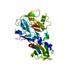
| ||||||||||||
|---|---|---|---|---|---|---|---|---|---|---|---|---|---|
| 1 |
| ||||||||||||
| Unit cell |
|
-Components
| #1: Protein | Mass: 39392.902 Da / Num. of mol.: 1 / Mutation: T64A,T233A,S333A,S334A Source method: isolated from a genetically manipulated source Source: (gene. exp.)  Homo sapiens (human) / References: UniProt: Q7KQK5, UniProt: Q8IKV6 Homo sapiens (human) / References: UniProt: Q7KQK5, UniProt: Q8IKV6 |
|---|---|
| #2: Water | ChemComp-HOH /  Mass: 18.015 Da / Num. of mol.: 100 / Source method: isolated from a natural source / Formula: H2O Mass: 18.015 Da / Num. of mol.: 100 / Source method: isolated from a natural source / Formula: H2O |
| Has protein modification | Y |
-Experimental details
-Experiment
| Experiment | Method: X-RAY DIFFRACTION / Number of used crystals: 1 |
|---|
- Sample preparation
| Crystal | Density Matthews: 1.89 Å3/Da / Density % sol: 36.03 % |
|---|---|
| Crystal grow | Temperature: 291.15 K / Method: vapor diffusion, hanging drop Details: 0.2 M Magnesium chloride, 0.1 M Tris (pH 8.5), 20 % (w/v) PEG 8000 |
-Data collection
| Diffraction | Mean temperature: 100 K / Serial crystal experiment: N |
|---|---|
| Diffraction source | Source: SYNCHROTRON / Site: APS / Beamline: 22-ID / Wavelength: 1 Å |
| Detector | Type: DECTRIS EIGER X 16M / Detector: PIXEL / Date: Apr 2, 2021 |
| Radiation | Protocol: SINGLE WAVELENGTH / Monochromatic (M) / Laue (L): M / Scattering type: x-ray |
| Radiation wavelength | Wavelength: 1 Å / Relative weight: 1 |
| Reflection | Resolution: 2.1→19.71 Å / Num. obs: 17236 / % possible obs: 98.4 % / Redundancy: 3.2 % / Biso Wilson estimate: 27.93 Å2 / CC1/2: 0.994 / CC star: 0.999 / Rmerge(I) obs: 0.0686 / Rpim(I) all: 0.0445 / Rrim(I) all: 0.082 / Net I/σ(I): 12.07 |
| Reflection shell | Resolution: 2.101→2.176 Å / Redundancy: 2.8 % / Rmerge(I) obs: 0.1663 / Mean I/σ(I) obs: 5.41 / Num. unique obs: 1600 / CC1/2: 0.953 / CC star: 0.988 / Rpim(I) all: 0.113 / Rrim(I) all: 0.2019 / % possible all: 92.38 |
- Processing
| Software |
| |||||||||||||||||||||||||||||||||||||||||||||||||
|---|---|---|---|---|---|---|---|---|---|---|---|---|---|---|---|---|---|---|---|---|---|---|---|---|---|---|---|---|---|---|---|---|---|---|---|---|---|---|---|---|---|---|---|---|---|---|---|---|---|---|
| Refinement | Method to determine structure: MOLECULAR REPLACEMENT / Resolution: 2.1→19.71 Å / SU ML: 0.2234 / Cross valid method: FREE R-VALUE / σ(F): 1.44 / Phase error: 21.3225 Stereochemistry target values: GeoStd + Monomer Library + CDL v1.2
| |||||||||||||||||||||||||||||||||||||||||||||||||
| Solvent computation | Shrinkage radii: 0.9 Å / VDW probe radii: 1.1 Å / Solvent model: FLAT BULK SOLVENT MODEL | |||||||||||||||||||||||||||||||||||||||||||||||||
| Displacement parameters | Biso mean: 34.6 Å2 | |||||||||||||||||||||||||||||||||||||||||||||||||
| Refinement step | Cycle: LAST / Resolution: 2.1→19.71 Å
| |||||||||||||||||||||||||||||||||||||||||||||||||
| Refine LS restraints |
| |||||||||||||||||||||||||||||||||||||||||||||||||
| LS refinement shell |
| |||||||||||||||||||||||||||||||||||||||||||||||||
| Refinement TLS params. | Method: refined / Origin x: 11.3207581173 Å / Origin y: 10.413349248 Å / Origin z: 5.03661866507 Å
| |||||||||||||||||||||||||||||||||||||||||||||||||
| Refinement TLS group | Selection details: all |
