 Movie
Movie Controller

[English] 日本語
 Yorodumi
- PDB-8gey: Crystal structure of human cellular retinol binding protein 1 in ... -
+ Open data

- Basic information
| Entry | Database: PDB / ID: 8gey | |||||||||
|---|---|---|---|---|---|---|---|---|---|---|
| Title | Crystal structure of human cellular retinol binding protein 1 in complex with 4-(hydroxymethyl)-1-[(4-methoxy-5,6,7,8-tetrahydronaphthalen-1-yl)sulfonyl]piperidin-4-ol | |||||||||
 Components Components | Retinol-binding protein 1 | |||||||||
 Keywords Keywords | Retinol-binding protein / RBP1 / CRBP1 / retinol-binding / retinol / vitamin A | |||||||||
| Function / homology |  Function and homology information Function and homology informationDefective visual phototransduction due to LRAT loss of function / all-trans-retinol binding / retinoic acid biosynthetic process / vitamin A metabolic process / retinoid binding / retinal binding / The canonical retinoid cycle in rods (twilight vision) / lipid homeostasis / fatty acid transport / Retinoid metabolism and transport ...Defective visual phototransduction due to LRAT loss of function / all-trans-retinol binding / retinoic acid biosynthetic process / vitamin A metabolic process / retinoid binding / retinal binding / The canonical retinoid cycle in rods (twilight vision) / lipid homeostasis / fatty acid transport / Retinoid metabolism and transport / lipid droplet / fatty acid binding / nucleoplasm / nucleus / cytosol Similarity search - Function | |||||||||
| Biological species |  Homo sapiens (human) Homo sapiens (human) | |||||||||
| Method |  X-RAY DIFFRACTION / SYNCHROTRON / MOLECULAR REPLACEMENT / Resolution: 1.3 Å X-RAY DIFFRACTION / SYNCHROTRON / MOLECULAR REPLACEMENT / Resolution: 1.3 Å | |||||||||
 Authors Authors | Plau, J. / Golczak, M. | |||||||||
| Funding support |  United States, 2items United States, 2items
| |||||||||
 Citation Citation | Journal: Acs Chem.Biol. / Year: 2023 Title: Discovery of Nonretinoid Inhibitors of CRBP1: Structural and Dynamic Insights for Ligand-Binding Mechanisms. Authors: Plau, J. / Morgan, C.E. / Fedorov, Y. / Banerjee, S. / Adams, D.J. / Blaner, W.S. / Yu, E.W. / Golczak, M. | |||||||||
| History |
|
- Structure visualization
| Structure viewer | Molecule: MolmilJmol/JSmol |
|---|
- Downloads & links
-Download
| PDBx/mmCIF format | 8gey.cif.gz | 95.5 KB | Display | PDBx/mmCIF format |
|---|---|---|---|---|
| PDB format | pdb8gey.ent.gz | 57.7 KB | Display | PDB format |
| PDBx/mmJSON format | 8gey.json.gz | Tree view | PDBx/mmJSON format | |
| Others |  Other downloads Other downloads |
-Validation report
| Summary document | 8gey_validation.pdf.gz | 742.4 KB | Display | wwPDB validaton report |
|---|---|---|---|---|
| Full document | 8gey_full_validation.pdf.gz | 742.9 KB | Display | |
| Data in XML | 8gey_validation.xml.gz | 11.6 KB | Display | |
| Data in CIF | 8gey_validation.cif.gz | 17.8 KB | Display | |
| Arichive directory | https://data.pdbj.org/pub/pdb/validation_reports/ge/8geyftp://data.pdbj.org/pub/pdb/validation_reports/ge/8gey | HTTPS FTP |
-Related structure data
| Related structure data | 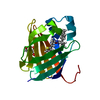 8gd2C 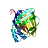 8gdmC 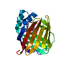 8gemC 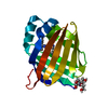 8geuC 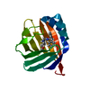 8gevC 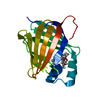 6e5lS C: citing same article ( S: Starting model for refinement |
|---|---|
| Similar structure data |
-Links
 PDBj
PDBj


- Assembly
| Deposited unit | 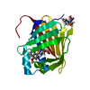
| ||||||||||||
|---|---|---|---|---|---|---|---|---|---|---|---|---|---|
| 1 |
| ||||||||||||
| Unit cell |
|
-Components
| #1: Protein | Mass: 16700.064 Da / Num. of mol.: 1 Source method: isolated from a genetically manipulated source Source: (gene. exp.) Homo sapiens (human) / Gene: RBP1, CRBP1 / Production host:  |
|---|---|
| #2: Chemical | ChemComp-ZE2 /   Mass: 355.449 Da / Num. of mol.: 1 / Source method: obtained synthetically / Formula: C17H25NO5S / Feature type: SUBJECT OF INVESTIGATION Mass: 355.449 Da / Num. of mol.: 1 / Source method: obtained synthetically / Formula: C17H25NO5S / Feature type: SUBJECT OF INVESTIGATION |
| #3: Chemical | ChemComp-BTB /   Mass: 209.240 Da / Num. of mol.: 1 / Source method: obtained synthetically / Formula: C8H19NO5 / Comment: pH buffer*YM Mass: 209.240 Da / Num. of mol.: 1 / Source method: obtained synthetically / Formula: C8H19NO5 / Comment: pH buffer*YM |
| #4: Water | ChemComp-HOH /  Mass: 18.015 Da / Num. of mol.: 313 / Source method: isolated from a natural source / Formula: H2O Mass: 18.015 Da / Num. of mol.: 313 / Source method: isolated from a natural source / Formula: H2O |
| Has ligand of interest | Y |
-Experimental details
-Experiment
| Experiment | Method: X-RAY DIFFRACTION / Number of used crystals: 1 |
|---|
- Sample preparation
| Crystal | Density Matthews: 2.15 Å3/Da / Density % sol: 42.88 % |
|---|---|
| Crystal grow | Temperature: 293 K / Method: vapor diffusion, sitting drop / pH: 5.5 Details: 0.1 M Bis-Tris, pH 5.5 and 25% poly ethylene glycol 3350 (w/v) |
-Data collection
| Diffraction | Mean temperature: 80 K / Serial crystal experiment: N |
|---|---|
| Diffraction source | Source: SYNCHROTRON / Site: APS / Beamline: 24-ID-C / Wavelength: 0.9791 Å |
| Detector | Type: DECTRIS PILATUS 6M-F / Detector: PIXEL / Date: Jun 20, 2020 |
| Radiation | Protocol: SINGLE WAVELENGTH / Monochromatic (M) / Laue (L): M / Scattering type: x-ray |
| Radiation wavelength | Wavelength: 0.9791 Å / Relative weight: 1 |
| Reflection | Resolution: 1.3→43.02 Å / Num. obs: 36077 / % possible obs: 99.4 % / Redundancy: 5.7 % / Biso Wilson estimate: 11.12 Å2 / CC1/2: 0.996 / Rmerge(I) obs: 0.091 / Rpim(I) all: 0.045 / Rrim(I) all: 0.11 / Net I/σ(I): 11.4 |
| Reflection shell | Resolution: 1.3→1.32 Å / Rmerge(I) obs: 0.755 / Mean I/σ(I) obs: 2 / Num. unique obs: 1765 / CC1/2: 0.67 / Rpim(I) all: 0.512 / Rrim(I) all: 0.641 / % possible all: 96.9 |
- Processing
| Software |
| ||||||||||||||||||||||||||||||||||||||||||||||||||||||||||||||||||||||||||||||||||||||||||||||||||
|---|---|---|---|---|---|---|---|---|---|---|---|---|---|---|---|---|---|---|---|---|---|---|---|---|---|---|---|---|---|---|---|---|---|---|---|---|---|---|---|---|---|---|---|---|---|---|---|---|---|---|---|---|---|---|---|---|---|---|---|---|---|---|---|---|---|---|---|---|---|---|---|---|---|---|---|---|---|---|---|---|---|---|---|---|---|---|---|---|---|---|---|---|---|---|---|---|---|---|---|
| Refinement | Method to determine structure: MOLECULAR REPLACEMENT Starting model: 6E5L Resolution: 1.3→38.75 Å / SU ML: 0.1026 / Cross valid method: FREE R-VALUE / σ(F): 1.33 / Phase error: 18.1743 Stereochemistry target values: GeoStd + Monomer Library + CDL v1.2
| ||||||||||||||||||||||||||||||||||||||||||||||||||||||||||||||||||||||||||||||||||||||||||||||||||
| Solvent computation | Shrinkage radii: 0.9 Å / VDW probe radii: 1.11 Å / Solvent model: FLAT BULK SOLVENT MODEL | ||||||||||||||||||||||||||||||||||||||||||||||||||||||||||||||||||||||||||||||||||||||||||||||||||
| Displacement parameters | Biso mean: 15.84 Å2 | ||||||||||||||||||||||||||||||||||||||||||||||||||||||||||||||||||||||||||||||||||||||||||||||||||
| Refinement step | Cycle: LAST / Resolution: 1.3→38.75 Å
| ||||||||||||||||||||||||||||||||||||||||||||||||||||||||||||||||||||||||||||||||||||||||||||||||||
| Refine LS restraints |
| ||||||||||||||||||||||||||||||||||||||||||||||||||||||||||||||||||||||||||||||||||||||||||||||||||
| LS refinement shell |
|
