 Movie
Movie Controller

[English] 日本語
 Yorodumi
- PDB-8gcv: XFEL structure of Mycobacterium tuberculosis beta lactamase micro... -
+ Open data

- Basic information
| Entry | Database: PDB / ID: 8gcv | ||||||
|---|---|---|---|---|---|---|---|
| Title | XFEL structure of Mycobacterium tuberculosis beta lactamase microcrystals | ||||||
 Components Components | Beta-lactamase | ||||||
 Keywords Keywords | HYDROLASE/Inhibitor / Beta lactamase / sulbactam / inhibitor / HYDROLASE / HYDROLASE-Inhibitor complex | ||||||
| Function / homology |  Function and homology information Function and homology informationbeta-lactam antibiotic catabolic process / beta-lactamase activity / beta-lactamase / periplasmic space / response to antibiotic / extracellular region Similarity search - Function | ||||||
| Biological species |   Mycobacterium tuberculosis (bacteria) Mycobacterium tuberculosis (bacteria) | ||||||
| Method |  X-RAY DIFFRACTION / FREE ELECTRON LASER / MOLECULAR REPLACEMENT / Resolution: 2.2 Å X-RAY DIFFRACTION / FREE ELECTRON LASER / MOLECULAR REPLACEMENT / Resolution: 2.2 Å | ||||||
 Authors Authors | Malla, T.N. / Schmidt, M. | ||||||
| Funding support |  United States, 1items United States, 1items
| ||||||
 Citation Citation | Journal: Nat Commun / Year: 2023 Title: Heterogeneity in M. tuberculosis beta-lactamase inhibition by Sulbactam. Authors: Malla, T.N. / Zielinski, K. / Aldama, L. / Bajt, S. / Feliz, D. / Hayes, B. / Hunter, M. / Kupitz, C. / Lisova, S. / Knoska, J. / Martin-Garcia, J.M. / Mariani, V. / Pandey, S. / Poudyal, I. ...Authors: Malla, T.N. / Zielinski, K. / Aldama, L. / Bajt, S. / Feliz, D. / Hayes, B. / Hunter, M. / Kupitz, C. / Lisova, S. / Knoska, J. / Martin-Garcia, J.M. / Mariani, V. / Pandey, S. / Poudyal, I. / Sierra, R.G. / Tolstikova, A. / Yefanov, O. / Yoon, C.H. / Ourmazd, A. / Fromme, P. / Schwander, P. / Barty, A. / Chapman, H.N. / Stojkovic, E.A. / Batyuk, A. / Boutet, S. / Phillips Jr., G.N. / Pollack, L. / Schmidt, M. | ||||||
| History |
|
- Structure visualization
| Structure viewer | Molecule: MolmilJmol/JSmol |
|---|
- Downloads & links
-Download
| PDBx/mmCIF format | 8gcv.cif.gz | 254.3 KB | Display | PDBx/mmCIF format |
|---|---|---|---|---|
| PDB format | pdb8gcv.ent.gz | 174.4 KB | Display | PDB format |
| PDBx/mmJSON format | 8gcv.json.gz | Tree view | PDBx/mmJSON format | |
| Others |  Other downloads Other downloads |
-Validation report
| Arichive directory | https://data.pdbj.org/pub/pdb/validation_reports/gc/8gcvftp://data.pdbj.org/pub/pdb/validation_reports/gc/8gcv | HTTPS FTP |
|---|
-Related structure data
| Related structure data | 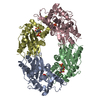 8ebiC  8ebrC 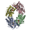 8ec4C 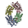 8ecfC 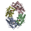 8gcsC 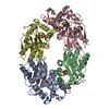 8gctC 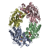 8gcxC 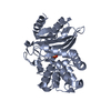 6b5xS C: citing same article ( S: Starting model for refinement |
|---|---|
| Similar structure data |
-Links
 PDBj
PDBj


- Assembly
| Deposited unit | 
| |||||||||||||||||||||||||||||||||||||||||||||||||||||||||||||||||||||||||||||||||||||||||||||||||||||||||||||||||||||
|---|---|---|---|---|---|---|---|---|---|---|---|---|---|---|---|---|---|---|---|---|---|---|---|---|---|---|---|---|---|---|---|---|---|---|---|---|---|---|---|---|---|---|---|---|---|---|---|---|---|---|---|---|---|---|---|---|---|---|---|---|---|---|---|---|---|---|---|---|---|---|---|---|---|---|---|---|---|---|---|---|---|---|---|---|---|---|---|---|---|---|---|---|---|---|---|---|---|---|---|---|---|---|---|---|---|---|---|---|---|---|---|---|---|---|---|---|---|---|
| 1 |
| |||||||||||||||||||||||||||||||||||||||||||||||||||||||||||||||||||||||||||||||||||||||||||||||||||||||||||||||||||||
| Unit cell |
| |||||||||||||||||||||||||||||||||||||||||||||||||||||||||||||||||||||||||||||||||||||||||||||||||||||||||||||||||||||
| Noncrystallographic symmetry (NCS) | NCS domain:
NCS domain segments:
|