[English] 日本語
 Yorodumi
- PDB-8gbr: Cardiac amyloid fibrils extracted from a wild-type ATTR amyloidos... -
+ Open data
- Basic information
| Entry | Database: PDB / ID: 8gbr | |||||||||
|---|---|---|---|---|---|---|---|---|---|---|
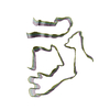
| Title | Cardiac amyloid fibrils extracted from a wild-type ATTR amyloidosis patient | |||||||||
 Components Components | Transthyretin | |||||||||
 Keywords Keywords | PROTEIN FIBRIL / Transthyretin / Amyloidosis / Systemic amyloidosis / ATTR / Cardiac | |||||||||
| Function / homology |  Function and homology information Function and homology informationDefective visual phototransduction due to STRA6 loss of function / negative regulation of glomerular filtration / The canonical retinoid cycle in rods (twilight vision) / hormone binding / purine nucleobase metabolic process / Non-integrin membrane-ECM interactions / molecular sequestering activity / phototransduction, visible light / retinoid metabolic process / Retinoid metabolism and transport ...Defective visual phototransduction due to STRA6 loss of function / negative regulation of glomerular filtration / The canonical retinoid cycle in rods (twilight vision) / hormone binding / purine nucleobase metabolic process / Non-integrin membrane-ECM interactions / molecular sequestering activity / phototransduction, visible light / retinoid metabolic process / Retinoid metabolism and transport / hormone activity / azurophil granule lumen / Amyloid fiber formation / Neutrophil degranulation / protein-containing complex binding / protein-containing complex / extracellular space / extracellular exosome / extracellular region / identical protein binding Similarity search - Function | |||||||||
| Biological species |  Homo sapiens (human) Homo sapiens (human) | |||||||||
| Method | ELECTRON MICROSCOPY / helical reconstruction / cryo EM / Resolution: 3.4 Å | |||||||||
 Authors Authors | Nguyen, B.A. / Saelices, L. | |||||||||
| Funding support |  United States, 2items United States, 2items
| |||||||||
 Citation Citation |  Journal: Commun Biol / Year: 2024 Journal: Commun Biol / Year: 2024Title: Cryo-EM confirms a common fibril fold in the heart of four patients with ATTRwt amyloidosis. Authors: Binh An Nguyen / Virender Singh / Shumaila Afrin / Preeti Singh / Maja Pekala / Yasmin Ahmed / Rose Pedretti / Jacob Canepa / Andrew Lemoff / Barbara Kluve-Beckerman / Pawel M Wydorski / ...Authors: Binh An Nguyen / Virender Singh / Shumaila Afrin / Preeti Singh / Maja Pekala / Yasmin Ahmed / Rose Pedretti / Jacob Canepa / Andrew Lemoff / Barbara Kluve-Beckerman / Pawel M Wydorski / Farzeen Chhapra / Lorena Saelices /  Abstract: ATTR amyloidosis results from the conversion of transthyretin into amyloid fibrils that deposit in tissues causing organ failure and death. This conversion is facilitated by mutations in ATTRv ...ATTR amyloidosis results from the conversion of transthyretin into amyloid fibrils that deposit in tissues causing organ failure and death. This conversion is facilitated by mutations in ATTRv amyloidosis, or aging in ATTRwt amyloidosis. ATTRv amyloidosis exhibits extreme phenotypic variability, whereas ATTRwt amyloidosis presentation is consistent and predictable. Previously, we found unique structural variabilities in cardiac amyloid fibrils from polyneuropathic ATTRv-I84S patients. In contrast, cardiac fibrils from five genotypically different patients with cardiomyopathy or mixed phenotypes are structurally homogeneous. To understand fibril structure's impact on phenotype, it is necessary to study the fibrils from multiple patients sharing genotype and phenotype. Here we show the cryo-electron microscopy structures of fibrils extracted from four cardiomyopathic ATTRwt amyloidosis patients. Our study confirms that they share identical conformations with minimal structural variability, consistent with their homogenous clinical presentation. Our study contributes to the understanding of ATTR amyloidosis biopathology and calls for further studies. | |||||||||
| History |
|
- Structure visualization
| Structure viewer | Molecule:  Molmil Molmil Jmol/JSmol Jmol/JSmol |
|---|
- Downloads & links
- Download
| PDBx/mmCIF format |  8gbr.cif.gz 8gbr.cif.gz | 101.7 KB | Display |  PDBx/mmCIF format PDBx/mmCIF format |
|---|---|---|---|---|
| PDB format |  pdb8gbr.ent.gz pdb8gbr.ent.gz | 75.9 KB | Display |  PDB format PDB format |
| PDBx/mmJSON format |  8gbr.json.gz 8gbr.json.gz | Tree view |  PDBx/mmJSON format PDBx/mmJSON format | |
| Others |  Other downloads Other downloads |
-Validation report
| Summary document |  8gbr_validation.pdf.gz 8gbr_validation.pdf.gz | 1 MB | Display |  wwPDB validaton report wwPDB validaton report |
|---|---|---|---|---|
| Full document |  8gbr_full_validation.pdf.gz 8gbr_full_validation.pdf.gz | 1 MB | Display | |
| Data in XML |  8gbr_validation.xml.gz 8gbr_validation.xml.gz | 26.7 KB | Display | |
| Data in CIF |  8gbr_validation.cif.gz 8gbr_validation.cif.gz | 38.2 KB | Display | |
| Arichive directory |  https://data.pdbj.org/pub/pdb/validation_reports/gb/8gbr https://data.pdbj.org/pub/pdb/validation_reports/gb/8gbr ftp://data.pdbj.org/pub/pdb/validation_reports/gb/8gbr ftp://data.pdbj.org/pub/pdb/validation_reports/gb/8gbr | HTTPS FTP |
-Related structure data
| Related structure data |  29920MC  8g9rC C: citing same article ( M: map data used to model this data |
|---|---|
| Similar structure data | Similarity search - Function & homology  F&H Search F&H Search |
- Links
Links
- Assembly
| Deposited unit | 
|
|---|---|
| 1 |
|
- Components
| #1: Protein | Mass: 15904.984 Da / Num. of mol.: 5 / Source method: isolated from a natural source / Source: (natural)  Homo sapiens (human) / Organ: Heart / Plasmid details: Transthyretin amyloidosis / Tissue: Cardiac / References: UniProt: P02766 Homo sapiens (human) / Organ: Heart / Plasmid details: Transthyretin amyloidosis / Tissue: Cardiac / References: UniProt: P02766 |
|---|
-Experimental details
-Experiment
| Experiment | Method: ELECTRON MICROSCOPY |
|---|---|
| EM experiment | Aggregation state: FILAMENT / 3D reconstruction method: helical reconstruction |
- Sample preparation
| Component | Name: cardiac amyloid fibril of wild-type transthyretin amyloidosis Type: TISSUE / Entity ID: all / Source: NATURAL |
|---|---|
| Source (natural) | Organism:  Homo sapiens (human) / Cellular location: extracellular / Organ: Heart / Tissue: Cardiac Homo sapiens (human) / Cellular location: extracellular / Organ: Heart / Tissue: Cardiac |
| Buffer solution | pH: 7 / Details: fibrils are in water |
| Specimen | Embedding applied: NO / Shadowing applied: NO / Staining applied: NO / Vitrification applied: YES / Details: Purified by water extraction |
| Specimen support | Grid material: COPPER / Grid mesh size: 300 divisions/in. / Grid type: Quantifoil R1.2/1.3 |
| Vitrification | Instrument: FEI VITROBOT MARK IV / Cryogen name: ETHANE / Humidity: 100 % / Chamber temperature: 295.15 K |
- Electron microscopy imaging
| Experimental equipment |  Model: Titan Krios / Image courtesy: FEI Company |
|---|---|
| Microscopy | Model: FEI TITAN KRIOS |
| Electron gun | Electron source:  FIELD EMISSION GUN / Accelerating voltage: 300 kV / Illumination mode: FLOOD BEAM FIELD EMISSION GUN / Accelerating voltage: 300 kV / Illumination mode: FLOOD BEAM |
| Electron lens | Mode: BRIGHT FIELD / Nominal defocus max: 2300 nm / Nominal defocus min: 1700 nm / Cs: 2.7 mm |
| Image recording | Average exposure time: 4.98 sec. / Electron dose: 40 e/Å2 / Film or detector model: GATAN K3 (6k x 4k) / Num. of grids imaged: 1 / Num. of real images: 8243 |
- Processing
| Software | Name: PHENIX / Version: 1.20.1_4487: / Classification: refinement | |||||||||||||||||||||||||||||||||||||||||||||
|---|---|---|---|---|---|---|---|---|---|---|---|---|---|---|---|---|---|---|---|---|---|---|---|---|---|---|---|---|---|---|---|---|---|---|---|---|---|---|---|---|---|---|---|---|---|---|
| EM software |
| |||||||||||||||||||||||||||||||||||||||||||||
| CTF correction | Type: NONE | |||||||||||||||||||||||||||||||||||||||||||||
| Helical symmerty | Angular rotation/subunit: -1.245 ° / Axial rise/subunit: 4.898 Å / Axial symmetry: C1 | |||||||||||||||||||||||||||||||||||||||||||||
| 3D reconstruction | Resolution: 3.4 Å / Resolution method: FSC 0.143 CUT-OFF / Num. of particles: 50584 / Num. of class averages: 1 / Symmetry type: HELICAL | |||||||||||||||||||||||||||||||||||||||||||||
| Atomic model building | B value: 71.63 / Protocol: RIGID BODY FIT / Space: REAL / Target criteria: Cross-correlation coefficient Details: Initial fitting was done using Coot with rigid body fit, then real space refinement for better fitting | |||||||||||||||||||||||||||||||||||||||||||||
| Atomic model building | PDB-ID: 8E7D Pdb chain-ID: A / Accession code: 8E7D / Details: Similar structure / Source name: PDB / Type: experimental model | |||||||||||||||||||||||||||||||||||||||||||||
| Refine LS restraints |
|
 Movie
Movie Controller


 PDBj
PDBj


