 Movie
Movie Controller

[English] 日本語
 Yorodumi
- PDB-8gba: Porous framework formed by assembly of a bipyridyl-conjugated hel... -
+ Open data

- Basic information
| Entry | Database: PDB / ID: 8gba | |||||||||
|---|---|---|---|---|---|---|---|---|---|---|
| Title | Porous framework formed by assembly of a bipyridyl-conjugated helical peptide | |||||||||
 Components Components | bipyridyl-conjugated helical peptide | |||||||||
 Keywords Keywords | DE NOVO PROTEIN | |||||||||
| Biological species | synthetic construct (others) | |||||||||
| Method |  X-RAY DIFFRACTION / SYNCHROTRON / MOLECULAR REPLACEMENT / Resolution: 0.89 Å X-RAY DIFFRACTION / SYNCHROTRON / MOLECULAR REPLACEMENT / Resolution: 0.89 Å | |||||||||
 Authors Authors | Hess, S.S. / Nguyen, A.I. | |||||||||
| Funding support | 1items
| |||||||||
 Citation Citation | Journal: J.Am.Chem.Soc. / Year: 2023 Title: Noncovalent Peptide Assembly Enables Crystalline, Permutable, and Reactive Thiol Frameworks. Authors: Hess, S.S. / Coppola, F. / Dang, V.T. / Tran, P.N. / Mickel, P.J. / Oktawiec, J. / Ren, Z. / Kral, P. / Nguyen, A.I. #1: Journal: Acta Crystallogr., Sect. D: Biol. Crystallogr. / Year: 2012Title: Towards automated crystallographic structure refinement with phenix.refine. Authors: Afonine, P.V. | |||||||||
| History |
|
- Structure visualization
| Structure viewer | Molecule:  MolmilJmol/JSmol MolmilJmol/JSmol |
|---|
- Downloads & links
-Download
| PDBx/mmCIF format | 8gba.cif.gz | 19 KB | Display | PDBx/mmCIF format |
|---|---|---|---|---|
| PDB format | pdb8gba.ent.gz | 10.8 KB | Display | PDB format |
| PDBx/mmJSON format | 8gba.json.gz | Tree view | PDBx/mmJSON format | |
| Others |  Other downloads Other downloads |
-Validation report
| Arichive directory | https://data.pdbj.org/pub/pdb/validation_reports/gb/8gbaftp://data.pdbj.org/pub/pdb/validation_reports/gb/8gba | HTTPS FTP |
|---|
-Related structure data
| Related structure data |  8gb9C 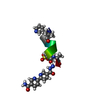 8gbhC  8gbiC  8gbmC 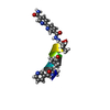 8gboC 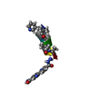 8gd6C 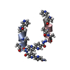 8gd8C 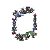 8givC 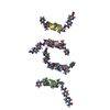 8gj7C 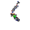 8gk1C  8gk2C 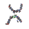 8gk9C 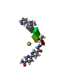 8gkbC 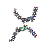 8gkxC 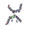 8gl0C  8gl4C 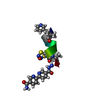 8gl5C 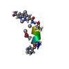 8sw2C 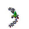 8sy4C C: citing same article ( |
|---|
-Links
 PDBj
PDBj
- Assembly
| Deposited unit | 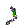
| ||||||||||||
|---|---|---|---|---|---|---|---|---|---|---|---|---|---|
| 1 |
| ||||||||||||
| Unit cell |
|
-Components
| #1: Protein/peptide | Mass: 1204.401 Da / Num. of mol.: 1 / Source method: obtained synthetically / Source: (synth.) synthetic construct (others) |
|---|---|
| #2: Water | ChemComp-HOH /  Mass: 18.015 Da / Num. of mol.: 12 / Source method: isolated from a natural source / Formula: H2O Mass: 18.015 Da / Num. of mol.: 12 / Source method: isolated from a natural source / Formula: H2O |
| Has ligand of interest | N |
-Experimental details
-Experiment
| Experiment | Method: X-RAY DIFFRACTION / Number of used crystals: 1 |
|---|
- Sample preparation
| Crystal | Density Matthews: 2.35 Å3/Da / Density % sol: 47.6 % |
|---|---|
| Crystal grow | Temperature: 298 K / Method: slow cooling / Details: Water/Acetonitrile |
-Data collection
| Diffraction | Mean temperature: 100 K / Serial crystal experiment: N |
|---|---|
| Diffraction source | Source: SYNCHROTRON / Site: APS  / Beamline: 21-ID-D / Wavelength: 0.61992 Å / Beamline: 21-ID-D / Wavelength: 0.61992 Å |
| Detector | Type: DECTRIS EIGER X 9M / Detector: PIXEL / Date: Mar 9, 2022 |
| Radiation | Protocol: SINGLE WAVELENGTH / Monochromatic (M) / Laue (L): M / Scattering type: x-ray |
| Radiation wavelength | Wavelength: 0.61992 Å / Relative weight: 1 |
| Reflection | Resolution: 0.89→28.63 Å / Num. obs: 11662 / % possible obs: 96.76 % / Redundancy: 12.4 % / Biso Wilson estimate: 3.52 Å2 / Rmerge(I) obs: 0.09684 / Net I/σ(I): 19.26 |
| Reflection shell | Resolution: 0.89→0.9219 Å / Rmerge(I) obs: 0.2003 / Num. unique obs: 547 |
- Processing
| Software |
| |||||||||||||||||||||||||||||||||||||||||||||||||||||||||||||||
|---|---|---|---|---|---|---|---|---|---|---|---|---|---|---|---|---|---|---|---|---|---|---|---|---|---|---|---|---|---|---|---|---|---|---|---|---|---|---|---|---|---|---|---|---|---|---|---|---|---|---|---|---|---|---|---|---|---|---|---|---|---|---|---|---|
| Refinement | Method to determine structure: MOLECULAR REPLACEMENT / Resolution: 0.89→28.63 Å / SU ML: 0.0433 / Cross valid method: FREE R-VALUE / σ(F): 1.37 / Phase error: 8.6222 Stereochemistry target values: GeoStd + Monomer Library + CDL v1.2
| |||||||||||||||||||||||||||||||||||||||||||||||||||||||||||||||
| Solvent computation | Shrinkage radii: 0.9 Å / VDW probe radii: 1.11 Å / Solvent model: FLAT BULK SOLVENT MODEL | |||||||||||||||||||||||||||||||||||||||||||||||||||||||||||||||
| Displacement parameters | Biso mean: 4.94 Å2 | |||||||||||||||||||||||||||||||||||||||||||||||||||||||||||||||
| Refinement step | Cycle: LAST / Resolution: 0.89→28.63 Å
| |||||||||||||||||||||||||||||||||||||||||||||||||||||||||||||||
| Refine LS restraints |
| |||||||||||||||||||||||||||||||||||||||||||||||||||||||||||||||
| LS refinement shell |
|
