 Movie
Movie Controller


+ Open data

- Basic information
| Entry | Database: PDB / ID: 8ga8 | |||||||||||||||||||||||||||||||||||||||
|---|---|---|---|---|---|---|---|---|---|---|---|---|---|---|---|---|---|---|---|---|---|---|---|---|---|---|---|---|---|---|---|---|---|---|---|---|---|---|---|---|
| Title | Structure of the yeast (HDAC) Rpd3L complex | |||||||||||||||||||||||||||||||||||||||
 Components Components |
| |||||||||||||||||||||||||||||||||||||||
 Keywords Keywords | TRANSCRIPTION / HDAC / Silencing / chromatin | |||||||||||||||||||||||||||||||||||||||
| Function / homology |  Function and homology information Function and homology informationnegative regulation of phosphatidylserine biosynthetic process / negative regulation of inositol biosynthetic process / positive regulation of phosphatidylserine biosynthetic process / negative regulation of phosphatidylcholine biosynthetic process / positive regulation of inositol biosynthetic process / regulation of invasive growth in response to glucose limitation / conjugation with cellular fusion / PI5P Regulates TP53 Acetylation / positive regulation of phosphatidylcholine biosynthetic process / Snt2C complex ...negative regulation of phosphatidylserine biosynthetic process / negative regulation of inositol biosynthetic process / positive regulation of phosphatidylserine biosynthetic process / negative regulation of phosphatidylcholine biosynthetic process / positive regulation of inositol biosynthetic process / regulation of invasive growth in response to glucose limitation / conjugation with cellular fusion / PI5P Regulates TP53 Acetylation / positive regulation of phosphatidylcholine biosynthetic process / Snt2C complex / negative regulation of silent mating-type cassette heterochromatin formation / positive regulation of invasive growth in response to glucose limitation / Rpd3L complex / protein localization to nucleolar rDNA repeats / negative regulation of reciprocal meiotic recombination / negative regulation of rDNA heterochromatin formation / Rpd3L-Expanded complex / invasive growth in response to glucose limitation / Rpd3S complex / rDNA chromatin condensation / nucleophagy / SUMOylation of transcription cofactors / HDACs deacetylate histones / histone deacetylase activity, hydrolytic mechanism / histone deacetylase / cell adhesion involved in single-species biofilm formation / histone H3K4me3 reader activity / histone deacetylase activity / SUMOylation of chromatin organization proteins / negative regulation of transcription by RNA polymerase I / cellular response to nitrogen starvation / regulation of DNA-templated DNA replication initiation / Sin3-type complex / histone deacetylase complex / positive regulation of macroautophagy / Estrogen-dependent gene expression / Ub-specific processing proteases / nuclear periphery / transcription coregulator activity / G1/S transition of mitotic cell cycle / transcription elongation by RNA polymerase II / meiotic cell cycle / double-strand break repair via nonhomologous end joining / G2/M transition of mitotic cell cycle / histone deacetylase binding / transcription corepressor activity / nucleosome assembly / cellular response to heat / heterochromatin formation / chromatin organization / response to oxidative stress / transcription coactivator activity / cell division / regulation of transcription by RNA polymerase II / regulation of DNA-templated transcription / chromatin / negative regulation of transcription by RNA polymerase II / positive regulation of transcription by RNA polymerase II / mitochondrion / zinc ion binding / identical protein binding / nucleus / cytosol / cytoplasm Similarity search - Function | |||||||||||||||||||||||||||||||||||||||
| Biological species |  | |||||||||||||||||||||||||||||||||||||||
| Method | ELECTRON MICROSCOPY / single particle reconstruction / cryo EM / Resolution: 3.5 Å | |||||||||||||||||||||||||||||||||||||||
 Authors Authors | Patel, A.B. / Radhakrishnan, I. / He, Y. | |||||||||||||||||||||||||||||||||||||||
| Funding support |  United States, 4items United States, 4items
| |||||||||||||||||||||||||||||||||||||||
 Citation Citation | Journal: Nat Commun / Year: 2023 Title: Cryo-EM structure of the Saccharomyces cerevisiae Rpd3L histone deacetylase complex. Authors: Avinash B Patel / Jinkang Qing / Kelly H Tam / Sara Zaman / Maria Luiso / Ishwar Radhakrishnan / Yuan He / Abstract: The Rpd3L histone deacetylase (HDAC) complex is an ancient 12-subunit complex conserved in a broad range of eukaryotes that performs localized deacetylation at or near sites of recruitment by DNA- ...The Rpd3L histone deacetylase (HDAC) complex is an ancient 12-subunit complex conserved in a broad range of eukaryotes that performs localized deacetylation at or near sites of recruitment by DNA-bound factors. Here we describe the cryo-EM structure of this prototypical HDAC complex that is characterized by as many as seven subunits performing scaffolding roles for the tight integration of the only catalytic subunit, Rpd3. The principal scaffolding protein, Sin3, along with Rpd3 and the histone chaperone, Ume1, are present in two copies, with each copy organized into separate lobes of an asymmetric dimeric molecular assembly. The active site of one Rpd3 is completely occluded by a leucine side chain of Rxt2, while the tips of the two lobes and the more peripherally associated subunits exhibit varying levels of flexibility and positional disorder. The structure reveals unexpected structural homology/analogy between unrelated subunits in the fungal and mammalian complexes and provides a foundation for deeper interrogations of structure, biology, and mechanism of these complexes, as well as for the discovery of HDAC complex-specific inhibitors. | |||||||||||||||||||||||||||||||||||||||
| History |
|
- Structure visualization
| Structure viewer | Molecule: MolmilJmol/JSmol |
|---|
- Downloads & links
-Download
| PDBx/mmCIF format | 8ga8.cif.gz | 546.9 KB | Display | PDBx/mmCIF format |
|---|---|---|---|---|
| PDB format | pdb8ga8.ent.gz | 409.8 KB | Display | PDB format |
| PDBx/mmJSON format | 8ga8.json.gz | Tree view | PDBx/mmJSON format | |
| Others |  Other downloads Other downloads |
-Validation report
| Arichive directory | https://data.pdbj.org/pub/pdb/validation_reports/ga/8ga8ftp://data.pdbj.org/pub/pdb/validation_reports/ga/8ga8 | HTTPS FTP |
|---|
-Related structure data
| Related structure data | 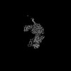 29892MC M: map data used to model this data C: citing same article ( |
|---|---|
| Similar structure data |


-Links
 PDBj
PDBj


- Assembly
| Deposited unit | 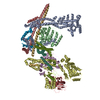
|
|---|---|
| 1 |
|
-Components
-Transcriptional regulatory protein ... , 7 types, 8 molecules HADJLMGK
| #1: Protein | Mass: 37685.301 Da / Num. of mol.: 1 / Source method: isolated from a natural source / Source: (natural) | ||||||||||
|---|---|---|---|---|---|---|---|---|---|---|---|
| #2: Protein | Mass: 175047.266 Da / Num. of mol.: 2 / Source method: isolated from a natural source / Source: (natural) #3: Protein | | Mass: 23064.215 Da / Num. of mol.: 1 / Source method: isolated from a natural source / Source: (natural) #4: Protein | | Mass: 37081.430 Da / Num. of mol.: 1 / Source method: isolated from a natural source / Source: (natural) #5: Protein | | Mass: 48684.082 Da / Num. of mol.: 1 / Source method: isolated from a natural source / Source: (natural) #6: Protein | | Mass: 47035.723 Da / Num. of mol.: 1 / Source method: isolated from a natural source / Source: (natural) #8: Protein | | Mass: 33851.535 Da / Num. of mol.: 1 / Source method: isolated from a natural source / Source: (natural) |
-Protein / Non-polymers , 2 types, 4 molecules BE
| #7: Protein | Mass: 48961.957 Da / Num. of mol.: 2 / Source method: isolated from a natural source / Source: (natural) #9: Chemical |  Mass: 65.409 Da / Num. of mol.: 2 / Source method: obtained synthetically / Formula: Zn / Feature type: SUBJECT OF INVESTIGATION Mass: 65.409 Da / Num. of mol.: 2 / Source method: obtained synthetically / Formula: Zn / Feature type: SUBJECT OF INVESTIGATION |
|---|
-Details
| Has ligand of interest | Y |
|---|---|
| Has protein modification | N |
-Experimental details
-Experiment
| Experiment | Method: ELECTRON MICROSCOPY |
|---|---|
| EM experiment | Aggregation state: PARTICLE / 3D reconstruction method: single particle reconstruction |
- Sample preparation
| Component | Name: Rpd3L / Type: COMPLEX / Entity ID: #1-#8 / Source: NATURAL |
|---|---|
| Source (natural) | Organism: |
| Buffer solution | pH: 7.9 |
| Specimen | Embedding applied: NO / Shadowing applied: NO / Staining applied: NO / Vitrification applied: YES |
| Specimen support | Grid material: COPPER / Grid mesh size: 300 divisions/in. / Grid type: Quantifoil R2/1 |
| Vitrification | Instrument: FEI VITROBOT MARK IV / Cryogen name: ETHANE / Humidity: 100 % / Chamber temperature: 277 K |
- Electron microscopy imaging
| Microscopy | Model: FEI TITAN |
|---|---|
| Electron gun | Electron source:  FIELD EMISSION GUN / Accelerating voltage: 300 kV / Illumination mode: FLOOD BEAM FIELD EMISSION GUN / Accelerating voltage: 300 kV / Illumination mode: FLOOD BEAM |
| Electron lens | Mode: BRIGHT FIELD / Nominal defocus max: 3000 nm / Nominal defocus min: 1000 nm |
| Image recording | Electron dose: 1 e/Å2 / Film or detector model: GATAN K3 (6k x 4k) |
- Processing
| Software | Name: PHENIX / Version: 1.20.1_4487: / Classification: refinement | ||||||||||||||||||||||||
|---|---|---|---|---|---|---|---|---|---|---|---|---|---|---|---|---|---|---|---|---|---|---|---|---|---|
| EM software |
| ||||||||||||||||||||||||
| CTF correction | Type: PHASE FLIPPING AND AMPLITUDE CORRECTION | ||||||||||||||||||||||||
| 3D reconstruction | Resolution: 3.5 Å / Resolution method: FSC 0.143 CUT-OFF / Num. of particles: 210993 / Symmetry type: POINT | ||||||||||||||||||||||||
| Refine LS restraints |
|
