 Movie
Movie Controller

[English] 日本語
 Yorodumi
- PDB-8g7i: Crystal Structure of FosB from Bacillus cereus with Zinc and Sulfate -
+ Open data

- Basic information
| Entry | Database: PDB / ID: 8g7i | ||||||
|---|---|---|---|---|---|---|---|
| Title | Crystal Structure of FosB from Bacillus cereus with Zinc and Sulfate | ||||||
 Components Components | Metallothiol transferase FosB | ||||||
 Keywords Keywords | TRANSFERASE / FosB / inhibitor / homodimer / bacillithiol-S-transferase | ||||||
| Function / homology |  Function and homology information Function and homology informationTransferases; Transferring alkyl or aryl groups, other than methyl groups / transferase activity, transferring alkyl or aryl (other than methyl) groups / response to antibiotic / magnesium ion binding / cytoplasm Similarity search - Function | ||||||
| Biological species |  | ||||||
| Method |  X-RAY DIFFRACTION / SYNCHROTRON / MOLECULAR REPLACEMENT / Resolution: 1.83 Å X-RAY DIFFRACTION / SYNCHROTRON / MOLECULAR REPLACEMENT / Resolution: 1.83 Å | ||||||
 Authors Authors | Travis, S. / Pang, A.H. / Tsodikov, O.V. / Garneau-Tsodikova, S. / Thompson, M.K. | ||||||
| Funding support |  United States, 1items United States, 1items
| ||||||
 Citation Citation | Journal: Rsc Med Chem / Year: 2023 Title: Identification and analysis of small molecule inhibitors of FosB from Staphylococcus aureus. Authors: Travis, S. / Green, K.D. / Thamban Chandrika, N. / Pang, A.H. / Frantom, P.A. / Tsodikov, O.V. / Garneau-Tsodikova, S. / Thompson, M.K. | ||||||
| History |
|
- Structure visualization
| Structure viewer | Molecule: MolmilJmol/JSmol |
|---|
- Downloads & links
-Download
| PDBx/mmCIF format | 8g7i.cif.gz | 80.6 KB | Display | PDBx/mmCIF format |
|---|---|---|---|---|
| PDB format | pdb8g7i.ent.gz | 57.8 KB | Display | PDB format |
| PDBx/mmJSON format | 8g7i.json.gz | Tree view | PDBx/mmJSON format | |
| Others |  Other downloads Other downloads |
-Validation report
| Arichive directory | https://data.pdbj.org/pub/pdb/validation_reports/g7/8g7iftp://data.pdbj.org/pub/pdb/validation_reports/g7/8g7i | HTTPS FTP |
|---|
-Related structure data


-Links
 PDBj
PDBj- Assembly
| Deposited unit | 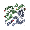
| ||||||||
|---|---|---|---|---|---|---|---|---|---|
| 1 |
| ||||||||
| Unit cell |
|
-Components
-Protein , 1 types, 2 molecules AB
| #1: Protein | Mass: 16488.484 Da / Num. of mol.: 2 Source method: isolated from a genetically manipulated source Source: (gene. exp.) References: UniProt: Q739M9, Transferases; Transferring alkyl or aryl groups, other than methyl groups |
|---|
-Non-polymers , 6 types, 270 molecules 


| #2: Chemical | ChemComp-FMT /  Mass: 46.025 Da / Num. of mol.: 5 / Source method: obtained synthetically / Formula: CH2O2 Mass: 46.025 Da / Num. of mol.: 5 / Source method: obtained synthetically / Formula: CH2O2#3: Chemical |  Mass: 24.305 Da / Num. of mol.: 2 / Source method: isolated from a natural source / Formula: Mg Mass: 24.305 Da / Num. of mol.: 2 / Source method: isolated from a natural source / Formula: Mg#4: Chemical |  Mass: 65.409 Da / Num. of mol.: 2 / Source method: isolated from a natural source / Formula: Zn Mass: 65.409 Da / Num. of mol.: 2 / Source method: isolated from a natural source / Formula: Zn#5: Chemical |  Mass: 96.063 Da / Num. of mol.: 2 / Source method: obtained synthetically / Formula: SO4 / Feature type: SUBJECT OF INVESTIGATION Mass: 96.063 Da / Num. of mol.: 2 / Source method: obtained synthetically / Formula: SO4 / Feature type: SUBJECT OF INVESTIGATION#6: Chemical | ChemComp-GOL / |  Mass: 92.094 Da / Num. of mol.: 1 / Source method: obtained synthetically / Formula: C3H8O3 Mass: 92.094 Da / Num. of mol.: 1 / Source method: obtained synthetically / Formula: C3H8O3#7: Water | ChemComp-HOH / | Mass: 18.015 Da / Num. of mol.: 258 / Source method: isolated from a natural source / Formula: H2O |
|---|
-Details
| Has ligand of interest | Y |
|---|
-Experimental details
-Experiment
| Experiment | Method: X-RAY DIFFRACTION / Number of used crystals: 1 |
|---|
- Sample preparation
| Crystal | Density Matthews: 2.29 Å3/Da / Density % sol: 46.36 % |
|---|---|
| Crystal grow | Temperature: 298 K / Method: vapor diffusion, hanging drop Details: Protein solution (10 mg/mL FosB in 20 mM HEPES buffer, pH 7.5 and 5 mM methylmalonate) and reservoir solution (50 mM Mg formate and 14% (w/v) PEG 3350) were mixed in a Hampton Research VDX ...Details: Protein solution (10 mg/mL FosB in 20 mM HEPES buffer, pH 7.5 and 5 mM methylmalonate) and reservoir solution (50 mM Mg formate and 14% (w/v) PEG 3350) were mixed in a Hampton Research VDX plate with a 2:3 ratio of protein:reservoir solutions. Cryoprotected in its reservoir solution with 20% glycerol added prior to rapid freezing in liquid nitrogen. |
-Data collection
| Diffraction | Mean temperature: 100 K / Serial crystal experiment: N | |||||||||||||||||||||||||||||||||||||||||||||||||||||||||||||||||||||||||||||||||||||||||||||||||||||||||||||||||||||||||||||||||||||||||||||||||||
|---|---|---|---|---|---|---|---|---|---|---|---|---|---|---|---|---|---|---|---|---|---|---|---|---|---|---|---|---|---|---|---|---|---|---|---|---|---|---|---|---|---|---|---|---|---|---|---|---|---|---|---|---|---|---|---|---|---|---|---|---|---|---|---|---|---|---|---|---|---|---|---|---|---|---|---|---|---|---|---|---|---|---|---|---|---|---|---|---|---|---|---|---|---|---|---|---|---|---|---|---|---|---|---|---|---|---|---|---|---|---|---|---|---|---|---|---|---|---|---|---|---|---|---|---|---|---|---|---|---|---|---|---|---|---|---|---|---|---|---|---|---|---|---|---|---|---|---|---|
| Diffraction source | Source: SYNCHROTRON / Site: APS / Beamline: 22-ID / Wavelength: 1 Å | |||||||||||||||||||||||||||||||||||||||||||||||||||||||||||||||||||||||||||||||||||||||||||||||||||||||||||||||||||||||||||||||||||||||||||||||||||
| Detector | Type: DECTRIS EIGER X 16M / Detector: PIXEL / Date: Sep 26, 2022 | |||||||||||||||||||||||||||||||||||||||||||||||||||||||||||||||||||||||||||||||||||||||||||||||||||||||||||||||||||||||||||||||||||||||||||||||||||
| Radiation | Protocol: SINGLE WAVELENGTH / Monochromatic (M) / Laue (L): M / Scattering type: x-ray | |||||||||||||||||||||||||||||||||||||||||||||||||||||||||||||||||||||||||||||||||||||||||||||||||||||||||||||||||||||||||||||||||||||||||||||||||||
| Radiation wavelength | Wavelength: 1 Å / Relative weight: 1 | |||||||||||||||||||||||||||||||||||||||||||||||||||||||||||||||||||||||||||||||||||||||||||||||||||||||||||||||||||||||||||||||||||||||||||||||||||
| Reflection | Resolution: 1.83→50 Å / Num. obs: 27845 / % possible obs: 99.8 % / Redundancy: 5.6 % / Rmerge(I) obs: 0.988 / Χ2: 0.062 / Net I/σ(I): 4.3 / Num. measured all: 296039 | |||||||||||||||||||||||||||||||||||||||||||||||||||||||||||||||||||||||||||||||||||||||||||||||||||||||||||||||||||||||||||||||||||||||||||||||||||
| Reflection shell |
|
- Processing
| Software |
| ||||||||||||||||||||||||||||||||||||||||||||||||||||||||||||||||||||||||||||||||||||||||||||||||||||||||||||||||||||||||||||||||||||||||||||||||||||||||||||||||||||||||||||||||||||||
|---|---|---|---|---|---|---|---|---|---|---|---|---|---|---|---|---|---|---|---|---|---|---|---|---|---|---|---|---|---|---|---|---|---|---|---|---|---|---|---|---|---|---|---|---|---|---|---|---|---|---|---|---|---|---|---|---|---|---|---|---|---|---|---|---|---|---|---|---|---|---|---|---|---|---|---|---|---|---|---|---|---|---|---|---|---|---|---|---|---|---|---|---|---|---|---|---|---|---|---|---|---|---|---|---|---|---|---|---|---|---|---|---|---|---|---|---|---|---|---|---|---|---|---|---|---|---|---|---|---|---|---|---|---|---|---|---|---|---|---|---|---|---|---|---|---|---|---|---|---|---|---|---|---|---|---|---|---|---|---|---|---|---|---|---|---|---|---|---|---|---|---|---|---|---|---|---|---|---|---|---|---|---|---|
| Refinement | Method to determine structure: MOLECULAR REPLACEMENT / Resolution: 1.83→48.94 Å / Cor.coef. Fo:Fc: 0.963 / Cor.coef. Fo:Fc free: 0.947 / Cross valid method: THROUGHOUT / ESU R: 0.121 / ESU R Free: 0.111 / Stereochemistry target values: MAXIMUM LIKELIHOOD / Details: HYDROGENS HAVE BEEN ADDED IN THE RIDING POSITIONS
| ||||||||||||||||||||||||||||||||||||||||||||||||||||||||||||||||||||||||||||||||||||||||||||||||||||||||||||||||||||||||||||||||||||||||||||||||||||||||||||||||||||||||||||||||||||||
| Solvent computation | Ion probe radii: 0.8 Å / Shrinkage radii: 0.8 Å / VDW probe radii: 1.2 Å / Solvent model: MASK | ||||||||||||||||||||||||||||||||||||||||||||||||||||||||||||||||||||||||||||||||||||||||||||||||||||||||||||||||||||||||||||||||||||||||||||||||||||||||||||||||||||||||||||||||||||||
| Displacement parameters | Biso mean: 17.88 Å2
| ||||||||||||||||||||||||||||||||||||||||||||||||||||||||||||||||||||||||||||||||||||||||||||||||||||||||||||||||||||||||||||||||||||||||||||||||||||||||||||||||||||||||||||||||||||||
| Refinement step | Cycle: 1 / Resolution: 1.83→48.94 Å
| ||||||||||||||||||||||||||||||||||||||||||||||||||||||||||||||||||||||||||||||||||||||||||||||||||||||||||||||||||||||||||||||||||||||||||||||||||||||||||||||||||||||||||||||||||||||
| Refine LS restraints |
|