 Movie
Movie Controller

[English] 日本語
 Yorodumi
- PDB-8g48: FphE, Staphylococcus aureus fluorophosphonate-binding serine hydr... -
+ Open data

- Basic information
| Entry | Database: PDB / ID: 8g48 | ||||||
|---|---|---|---|---|---|---|---|
| Title | FphE, Staphylococcus aureus fluorophosphonate-binding serine hydrolases E, dimeric apo form | ||||||
 Components Components | Fluorophosphonate-binding serine hydrolase E | ||||||
 Keywords Keywords | HYDROLASE / FphE / Staphylococcus aureus / S. aureus / fluorophosphonate-binding / serine hydrolases / lipase | ||||||
| Function / homology | Hydrolases / : / alpha/beta hydrolase fold / Alpha/beta hydrolase fold-1 / Alpha/Beta hydrolase fold / hydrolase activity / membrane / Uncharacterized hydrolase SAUSA300_2518 Function and homology information Function and homology information | ||||||
| Biological species |  Staphylococcus aureus USA300-CA-263 (bacteria) Staphylococcus aureus USA300-CA-263 (bacteria) | ||||||
| Method |  X-RAY DIFFRACTION / SYNCHROTRON / MOLECULAR REPLACEMENT / Resolution: 1.95 Å X-RAY DIFFRACTION / SYNCHROTRON / MOLECULAR REPLACEMENT / Resolution: 1.95 Å | ||||||
 Authors Authors | Fellner, M. | ||||||
| Funding support |  New Zealand, 1items New Zealand, 1items
| ||||||
 Citation Citation | Journal: To Be Published Title: FphE, Staphylococcus aureus fluorophosphonate-binding serine hydrolases E, dimeric apo form Authors: Fellner, M. | ||||||
| History |
|
- Structure visualization
| Structure viewer | Molecule: MolmilJmol/JSmol |
|---|
- Downloads & links
-Download
| PDBx/mmCIF format | 8g48.cif.gz | 125.1 KB | Display | PDBx/mmCIF format |
|---|---|---|---|---|
| PDB format | pdb8g48.ent.gz | 96.2 KB | Display | PDB format |
| PDBx/mmJSON format | 8g48.json.gz | Tree view | PDBx/mmJSON format | |
| Others |  Other downloads Other downloads |
-Validation report
| Arichive directory | https://data.pdbj.org/pub/pdb/validation_reports/g4/8g48ftp://data.pdbj.org/pub/pdb/validation_reports/g4/8g48 | HTTPS FTP |
|---|
-Related structure data
| Similar structure data |
|---|
-Links
 PDBj
PDBj


- Assembly
| Deposited unit | 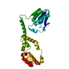
| ||||||||
|---|---|---|---|---|---|---|---|---|---|
| 1 | 
| ||||||||
| Unit cell |
|
-Components
| #1: Protein | Mass: 31275.100 Da / Num. of mol.: 1 Source method: isolated from a genetically manipulated source Source: (gene. exp.) Staphylococcus aureus USA300-CA-263 (bacteria)Gene: SAUSA300_2518 / Plasmid: F1010 / Production host: |
|---|---|
| #2: Water | ChemComp-HOH /  Mass: 18.015 Da / Num. of mol.: 137 / Source method: isolated from a natural source / Formula: H2O Mass: 18.015 Da / Num. of mol.: 137 / Source method: isolated from a natural source / Formula: H2O |
-Experimental details
-Experiment
| Experiment | Method: X-RAY DIFFRACTION / Number of used crystals: 1 |
|---|
- Sample preparation
| Crystal | Density Matthews: 2.19 Å3/Da / Density % sol: 43.92 % |
|---|---|
| Crystal grow | Temperature: 289.15 K / Method: vapor diffusion, sitting drop / pH: 8.5 Details: 0.3 ul 1.9 mg/ml FphE (10mM HEPES pH 7.6, 100mM NaCl) were mixed with 0.3 ul of reservoir solution. Sitting drop reservoir contained 25 ul of 200mM Magnesium chloride hexahydrate, 100mM Tris ...Details: 0.3 ul 1.9 mg/ml FphE (10mM HEPES pH 7.6, 100mM NaCl) were mixed with 0.3 ul of reservoir solution. Sitting drop reservoir contained 25 ul of 200mM Magnesium chloride hexahydrate, 100mM Tris pH 8.5, 25% PEG 2000 MME. Crystal appeared within ~30 days at 16C. It was frozen in a solution of ~25% glycerol, 75% reservoir. |
-Data collection
| Diffraction | Mean temperature: 100 K / Serial crystal experiment: N |
|---|---|
| Diffraction source | Source: SYNCHROTRON / Site: Australian Synchrotron  / Beamline: MX2 / Wavelength: 0.9537 Å / Beamline: MX2 / Wavelength: 0.9537 Å |
| Detector | Type: DECTRIS EIGER X 16M / Detector: PIXEL / Date: Jun 21, 2022 |
| Radiation | Protocol: SINGLE WAVELENGTH / Monochromatic (M) / Laue (L): M / Scattering type: x-ray |
| Radiation wavelength | Wavelength: 0.9537 Å / Relative weight: 1 |
| Reflection | Resolution: 1.95→41.94 Å / Num. obs: 20746 / % possible obs: 99.5 % / Redundancy: 6.1 % / CC1/2: 0.997 / Rmerge(I) obs: 0.098 / Rpim(I) all: 0.043 / Rrim(I) all: 0.107 / Χ2: 0.9 / Net I/σ(I): 9.7 / Num. measured all: 126180 |
| Reflection shell | Resolution: 1.95→2 Å / % possible obs: 95.2 % / Redundancy: 6 % / Rmerge(I) obs: 0.802 / Num. measured all: 8309 / Num. unique obs: 1384 / CC1/2: 0.717 / Rpim(I) all: 0.352 / Rrim(I) all: 0.878 / Χ2: 0.77 / Net I/σ(I) obs: 2.1 |
- Processing
| Software |
| ||||||||||||||||||||||||||||||||||||||||||||||||||||||||||||||||||||||||||||||||||||||||||||||||||||
|---|---|---|---|---|---|---|---|---|---|---|---|---|---|---|---|---|---|---|---|---|---|---|---|---|---|---|---|---|---|---|---|---|---|---|---|---|---|---|---|---|---|---|---|---|---|---|---|---|---|---|---|---|---|---|---|---|---|---|---|---|---|---|---|---|---|---|---|---|---|---|---|---|---|---|---|---|---|---|---|---|---|---|---|---|---|---|---|---|---|---|---|---|---|---|---|---|---|---|---|---|---|
| Refinement | Method to determine structure: MOLECULAR REPLACEMENT / Resolution: 1.95→41.94 Å / SU ML: 0.23 / Cross valid method: FREE R-VALUE / σ(F): 1.34 / Phase error: 23.34 / Stereochemistry target values: ML
| ||||||||||||||||||||||||||||||||||||||||||||||||||||||||||||||||||||||||||||||||||||||||||||||||||||
| Solvent computation | Shrinkage radii: 0.9 Å / VDW probe radii: 1.1 Å / Solvent model: FLAT BULK SOLVENT MODEL | ||||||||||||||||||||||||||||||||||||||||||||||||||||||||||||||||||||||||||||||||||||||||||||||||||||
| Refinement step | Cycle: LAST / Resolution: 1.95→41.94 Å
| ||||||||||||||||||||||||||||||||||||||||||||||||||||||||||||||||||||||||||||||||||||||||||||||||||||
| Refine LS restraints |
| ||||||||||||||||||||||||||||||||||||||||||||||||||||||||||||||||||||||||||||||||||||||||||||||||||||
| LS refinement shell |
| ||||||||||||||||||||||||||||||||||||||||||||||||||||||||||||||||||||||||||||||||||||||||||||||||||||
| Refinement TLS params. | Method: refined / Refine-ID: X-RAY DIFFRACTION
| ||||||||||||||||||||||||||||||||||||||||||||||||||||||||||||||||||||||||||||||||||||||||||||||||||||
| Refinement TLS group |
|
