 Movie
Movie Controller

[English] 日本語
 Yorodumi
- PDB-8g28: Crystal Structure of the C-terminal Fragment of AAA ATPase from S... -
+ Open data

- Basic information
| Entry | Database: PDB / ID: 8g28 | |||||||||
|---|---|---|---|---|---|---|---|---|---|---|
| Title | Crystal Structure of the C-terminal Fragment of AAA ATPase from Streptococcus pneumoniae. | |||||||||
 Components Components | ATPase, AAA family | |||||||||
 Keywords Keywords | HYDROLASE / Center for Structural Biology of Infectious Diseases (CSBID) / Structural Genomics / Center for Structural Genomics of Infectious Diseases / CSGID / AAA ATPase | |||||||||
| Function / homology |  Function and homology information Function and homology information | |||||||||
| Biological species |  Streptococcus pneumoniae TIGR4 (bacteria) Streptococcus pneumoniae TIGR4 (bacteria) | |||||||||
| Method |  X-RAY DIFFRACTION / SYNCHROTRON / SAD / Resolution: 1.3 Å X-RAY DIFFRACTION / SYNCHROTRON / SAD / Resolution: 1.3 Å | |||||||||
 Authors Authors | Minasov, G. / Shuvalova, L. / Brunzelle, J.S. / Kiryukhina, O. / Satchell, K.J.F. / Center for Structural Biology of Infectious Diseases (CSBID) / Center for Structural Genomics of Infectious Diseases (CSGID) | |||||||||
| Funding support |  United States, 2items United States, 2items
| |||||||||
 Citation Citation | Journal: To Be Published Title: Crystal Structure of the C-terminal Fragment of AAA ATPase from Streptococcus pneumoniae. Authors: Minasov, G. / Shuvalova, L. / Brunzelle, J.S. / Kiryukhina, O. / Satchell, K.J.F. / Center for Structural Biology of Infectious Diseases (CSBID) / Center for Structural Genomics of Infectious Diseases (CSGID) | |||||||||
| History |
|
- Structure visualization
| Structure viewer | Molecule: MolmilJmol/JSmol |
|---|
- Downloads & links
-Download
| PDBx/mmCIF format | 8g28.cif.gz | 134.1 KB | Display | PDBx/mmCIF format |
|---|---|---|---|---|
| PDB format | pdb8g28.ent.gz | 100.3 KB | Display | PDB format |
| PDBx/mmJSON format | 8g28.json.gz | Tree view | PDBx/mmJSON format | |
| Others |  Other downloads Other downloads |
-Validation report
| Summary document | 8g28_validation.pdf.gz | 428.2 KB | Display | wwPDB validaton report |
|---|---|---|---|---|
| Full document | 8g28_full_validation.pdf.gz | 429.8 KB | Display | |
| Data in XML | 8g28_validation.xml.gz | 14 KB | Display | |
| Data in CIF | 8g28_validation.cif.gz | 21.3 KB | Display | |
| Arichive directory | https://data.pdbj.org/pub/pdb/validation_reports/g2/8g28ftp://data.pdbj.org/pub/pdb/validation_reports/g2/8g28 | HTTPS FTP |
-Related structure data
| Similar structure data | |
|---|---|
| Other databases |
-Links
 PDBj
PDBj
- Assembly
| Deposited unit | 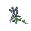
| ||||||||||||
|---|---|---|---|---|---|---|---|---|---|---|---|---|---|
| 1 | 
| ||||||||||||
| Unit cell |
| ||||||||||||
| Components on special symmetry positions |
|
-Components
| #1: Protein | Mass: 47706.754 Da / Num. of mol.: 2 Source method: isolated from a genetically manipulated source Details: The protein was hydrolyzed during the crystallization experiment. Source: (gene. exp.) Streptococcus pneumoniae TIGR4 (bacteria)Gene: SP_1790 / Plasmid: pMCSG53 / Production host: #2: Chemical | ChemComp-CL / |   Mass: 35.453 Da / Num. of mol.: 1 / Source method: obtained synthetically / Formula: Cl Mass: 35.453 Da / Num. of mol.: 1 / Source method: obtained synthetically / Formula: Cl#3: Water | ChemComp-HOH / |  Mass: 18.015 Da / Num. of mol.: 320 / Source method: isolated from a natural source / Formula: H2O Mass: 18.015 Da / Num. of mol.: 320 / Source method: isolated from a natural source / Formula: H2OHas ligand of interest | N | Has protein modification | Y | |
|---|
-Experimental details
-Experiment
| Experiment | Method: X-RAY DIFFRACTION / Number of used crystals: 1 |
|---|
- Sample preparation
| Crystal | Density Matthews: 2.3 Å3/Da / Density % sol: 46.6 % |
|---|---|
| Crystal grow | Temperature: 292 K / Method: vapor diffusion, sitting drop / pH: 7.5 Details: Protein: 5.6 mg/ml, 0.25M Sodium chloride, 0.01M Tris pH 8.3; Screen: Classics II (F12), 0.2M Sodium chloride, 0.1M HEPES pH 7.5, 25% (w/v) PEG3350. |
-Data collection
| Diffraction | Mean temperature: 100 K / Serial crystal experiment: N |
|---|---|
| Diffraction source | Source: SYNCHROTRON / Site: APS / Beamline: 21-ID-G / Wavelength: 0.9786 Å |
| Detector | Type: MARMOSAIC 300 mm CCD / Detector: CCD / Date: Jul 28, 2021 |
| Radiation | Monochromator: C(111) / Protocol: SINGLE WAVELENGTH / Monochromatic (M) / Laue (L): M / Scattering type: x-ray |
| Radiation wavelength | Wavelength: 0.9786 Å / Relative weight: 1 |
| Reflection | Resolution: 1.3→30 Å / Num. obs: 57219 / % possible obs: 99.9 % / Observed criterion σ(I): -3 / Redundancy: 24.4 % / Biso Wilson estimate: 13.3 Å2 / CC1/2: 0.999 / CC star: 1 / Rmerge(I) obs: 0.082 / Rpim(I) all: 0.017 / Rrim(I) all: 0.084 / Rsym value: 0.082 / Χ2: 1.085 / Net I/σ(I): 42.8 |
| Reflection shell | Resolution: 1.3→1.32 Å / Redundancy: 21.1 % / Rmerge(I) obs: 1.512 / Mean I/σ(I) obs: 2.4 / Num. unique obs: 2800 / CC1/2: 0.731 / CC star: 0.919 / Rpim(I) all: 0.331 / Rsym value: 1.512 / Χ2: 1.005 / % possible all: 100 |
- Processing
| Software |
| |||||||||||||||||||||||||||||||||||||||||||||||||||||||||||||||||||||||||||||||||||||||||||||||||||||||||||||||||||||||||||||||||||||||||||||||||||||||||||||||||||||||||||||||||||||||||||||||||||||||||||||||||||||||||||||||||||||||||||||||||||||||||||||||||||||||||||||||||||||||||||||||||||||||||||||||||||||||||||||||||||||
|---|---|---|---|---|---|---|---|---|---|---|---|---|---|---|---|---|---|---|---|---|---|---|---|---|---|---|---|---|---|---|---|---|---|---|---|---|---|---|---|---|---|---|---|---|---|---|---|---|---|---|---|---|---|---|---|---|---|---|---|---|---|---|---|---|---|---|---|---|---|---|---|---|---|---|---|---|---|---|---|---|---|---|---|---|---|---|---|---|---|---|---|---|---|---|---|---|---|---|---|---|---|---|---|---|---|---|---|---|---|---|---|---|---|---|---|---|---|---|---|---|---|---|---|---|---|---|---|---|---|---|---|---|---|---|---|---|---|---|---|---|---|---|---|---|---|---|---|---|---|---|---|---|---|---|---|---|---|---|---|---|---|---|---|---|---|---|---|---|---|---|---|---|---|---|---|---|---|---|---|---|---|---|---|---|---|---|---|---|---|---|---|---|---|---|---|---|---|---|---|---|---|---|---|---|---|---|---|---|---|---|---|---|---|---|---|---|---|---|---|---|---|---|---|---|---|---|---|---|---|---|---|---|---|---|---|---|---|---|---|---|---|---|---|---|---|---|---|---|---|---|---|---|---|---|---|---|---|---|---|---|---|---|---|---|---|---|---|---|---|---|---|---|---|---|---|---|---|---|---|---|---|---|---|---|---|---|---|---|---|---|---|---|---|---|---|---|---|---|---|---|---|---|---|---|---|---|---|---|---|---|---|---|---|---|---|---|---|---|---|---|---|---|---|---|---|---|
| Refinement | Method to determine structure: SAD / Resolution: 1.3→29.18 Å / Cor.coef. Fo:Fc: 0.98 / Cor.coef. Fo:Fc free: 0.975 / SU B: 1.12 / SU ML: 0.021 / Cross valid method: THROUGHOUT / σ(F): 0 / ESU R: 0.041 / ESU R Free: 0.039 / Stereochemistry target values: MAXIMUM LIKELIHOOD Details: HYDROGENS HAVE BEEN ADDED IN THE RIDING POSITIONS U VALUES : WITH TLS ADDED
| |||||||||||||||||||||||||||||||||||||||||||||||||||||||||||||||||||||||||||||||||||||||||||||||||||||||||||||||||||||||||||||||||||||||||||||||||||||||||||||||||||||||||||||||||||||||||||||||||||||||||||||||||||||||||||||||||||||||||||||||||||||||||||||||||||||||||||||||||||||||||||||||||||||||||||||||||||||||||||||||||||||
| Solvent computation | Ion probe radii: 0.8 Å / Shrinkage radii: 0.8 Å / VDW probe radii: 1.2 Å / Solvent model: MASK | |||||||||||||||||||||||||||||||||||||||||||||||||||||||||||||||||||||||||||||||||||||||||||||||||||||||||||||||||||||||||||||||||||||||||||||||||||||||||||||||||||||||||||||||||||||||||||||||||||||||||||||||||||||||||||||||||||||||||||||||||||||||||||||||||||||||||||||||||||||||||||||||||||||||||||||||||||||||||||||||||||||
| Displacement parameters | Biso max: 81.3 Å2 / Biso mean: 16.748 Å2 / Biso min: 8.4 Å2
| |||||||||||||||||||||||||||||||||||||||||||||||||||||||||||||||||||||||||||||||||||||||||||||||||||||||||||||||||||||||||||||||||||||||||||||||||||||||||||||||||||||||||||||||||||||||||||||||||||||||||||||||||||||||||||||||||||||||||||||||||||||||||||||||||||||||||||||||||||||||||||||||||||||||||||||||||||||||||||||||||||||
| Refinement step | Cycle: final / Resolution: 1.3→29.18 Å
| |||||||||||||||||||||||||||||||||||||||||||||||||||||||||||||||||||||||||||||||||||||||||||||||||||||||||||||||||||||||||||||||||||||||||||||||||||||||||||||||||||||||||||||||||||||||||||||||||||||||||||||||||||||||||||||||||||||||||||||||||||||||||||||||||||||||||||||||||||||||||||||||||||||||||||||||||||||||||||||||||||||
| Refine LS restraints |
| |||||||||||||||||||||||||||||||||||||||||||||||||||||||||||||||||||||||||||||||||||||||||||||||||||||||||||||||||||||||||||||||||||||||||||||||||||||||||||||||||||||||||||||||||||||||||||||||||||||||||||||||||||||||||||||||||||||||||||||||||||||||||||||||||||||||||||||||||||||||||||||||||||||||||||||||||||||||||||||||||||||
| LS refinement shell | Resolution: 1.3→1.334 Å / Rfactor Rfree error: 0 / Total num. of bins used: 20
| |||||||||||||||||||||||||||||||||||||||||||||||||||||||||||||||||||||||||||||||||||||||||||||||||||||||||||||||||||||||||||||||||||||||||||||||||||||||||||||||||||||||||||||||||||||||||||||||||||||||||||||||||||||||||||||||||||||||||||||||||||||||||||||||||||||||||||||||||||||||||||||||||||||||||||||||||||||||||||||||||||||
| Refinement TLS params. | Method: refined / Refine-ID: X-RAY DIFFRACTION
| |||||||||||||||||||||||||||||||||||||||||||||||||||||||||||||||||||||||||||||||||||||||||||||||||||||||||||||||||||||||||||||||||||||||||||||||||||||||||||||||||||||||||||||||||||||||||||||||||||||||||||||||||||||||||||||||||||||||||||||||||||||||||||||||||||||||||||||||||||||||||||||||||||||||||||||||||||||||||||||||||||||
| Refinement TLS group |
|
