 Movie
Movie Controller

[English] 日本語
 Yorodumi
- PDB-8g1r: A Vibrio cholerae viral satellite enables efficient horizontal tr... -
+ Open data

- Basic information
| Entry | Database: PDB / ID: 8g1r | |||||||||||||||||||||
|---|---|---|---|---|---|---|---|---|---|---|---|---|---|---|---|---|---|---|---|---|---|---|
| Title | A Vibrio cholerae viral satellite enables efficient horizontal transfer by using an external scaffold to assemble hijacked coat proteins into small capsids | |||||||||||||||||||||
 Components Components |
| |||||||||||||||||||||
 Keywords Keywords | VIRUS LIKE PARTICLE / PLE / Procapsid | |||||||||||||||||||||
| Function / homology | Major capsid protein GpE / Phage major capsid protein E / host cell cytoplasm / Putative major head protein / Uncharacterized protein Function and homology information Function and homology information | |||||||||||||||||||||
| Biological species |  Vibrio phage ICP1_2011_A (virus) Vibrio phage ICP1_2011_A (virus)  Vibrio cholerae (bacteria) Vibrio cholerae (bacteria) | |||||||||||||||||||||
| Method | ELECTRON MICROSCOPY / single particle reconstruction / cryo EM / Resolution: 3.4 Å | |||||||||||||||||||||
 Authors Authors | Subramanian, S. / Boyd, C.M. / Seed, K.D. / Parent, K.N. | |||||||||||||||||||||
| Funding support |  United States, 6items United States, 6items
| |||||||||||||||||||||
 Citation Citation | Journal: bioRxiv / Year: 2023 Title: A viral satellite maximizes its spread and inhibits phage by remodeling hijacked phage coat proteins into small capsids. Authors: Caroline M Boyd / Sundharraman Subramanian / Drew T Dunham / Kristin N Parent / Kimberley D Seed / Abstract: Phage satellites commonly remodel capsids they hijack from the phages they parasitize, but only a few mechanisms regulating the change in capsid size have been reported. Here, we investigated how a ...Phage satellites commonly remodel capsids they hijack from the phages they parasitize, but only a few mechanisms regulating the change in capsid size have been reported. Here, we investigated how a satellite from , PLE, remodels the capsid it has been predicted to steal from the phage ICP1 (1). We identified that a PLE-encoded protein, TcaP, is both necessary and sufficient to form small capsids during ICP1 infection. Interestingly, we found that PLE is dependent on small capsids for efficient transduction of its genome, making it the first satellite to have this requirement. ICP1 isolates that escaped TcaP-mediated remodeling acquired substitutions in the coat protein, suggesting an interaction between these two proteins. With a procapsid-like-particle (PLP) assembly platform in , we demonstrated that TcaP is a scaffold that regulates the assembly of small capsids. Further, we studied the structure of PLE PLPs using cryogenic electron microscopy and found that TcaP is an external scaffold, that is functionally and somewhat structurally similar to the external scaffold, Sid, encoded by the unrelated satellite P4 (2). Finally, we showed that TcaP is largely conserved across PLEs. Together, these data support a model in which TcaP directs the assembly of small capsids comprised of ICP1 coat proteins, which inhibits the complete packaging of the ICP1 genome and permits more efficient packaging of replicated PLE genomes. #1: Journal: Elife / Year: 2024 Title: A viral satellite maximizes its spread and inhibits phage by remodeling hijacked phage coat proteins into small capsids. Authors: Caroline M Boyd / Sundharraman Subramanian / Drew T Dunham / Kristin N Parent / Kimberley D Seed / Abstract: Phage satellites commonly remodel capsids they hijack from the phages they parasitize, but only a few mechanisms regulating the change in capsid size have been reported. Here, we investigated how a ...Phage satellites commonly remodel capsids they hijack from the phages they parasitize, but only a few mechanisms regulating the change in capsid size have been reported. Here, we investigated how a satellite from , phage-inducible chromosomal island-like element (PLE), remodels the capsid it has been predicted to steal from the phage ICP1 (Netter et al., 2021). We identified that a PLE-encoded protein, TcaP, is both necessary and sufficient to form small capsids during ICP1 infection. Interestingly, we found that PLE is dependent on small capsids for efficient transduction of its genome, making it the first satellite to have this requirement. ICP1 isolates that escaped TcaP-mediated remodeling acquired substitutions in the coat protein, suggesting an interaction between these two proteins. With a procapsid-like particle (PLP) assembly platform in , we demonstrated that TcaP is a bona fide scaffold that regulates the assembly of small capsids. Further, we studied the structure of PLE PLPs using cryogenic electron microscopy and found that TcaP is an external scaffold that is functionally and somewhat structurally similar to the external scaffold, Sid, encoded by the unrelated satellite P4 (Kizziah et al., 2020). Finally, we showed that TcaP is largely conserved across PLEs. Together, these data support a model in which TcaP directs the assembly of small capsids comprised of ICP1 coat proteins, which inhibits the complete packaging of the ICP1 genome and permits more efficient packaging of replicated PLE genomes. | |||||||||||||||||||||
| History |
|
- Structure visualization
| Structure viewer | Molecule: MolmilJmol/JSmol |
|---|
- Downloads & links
-Download
| PDBx/mmCIF format | 8g1r.cif.gz | 243 KB | Display | PDBx/mmCIF format |
|---|---|---|---|---|
| PDB format | pdb8g1r.ent.gz | 187.6 KB | Display | PDB format |
| PDBx/mmJSON format | 8g1r.json.gz | Tree view | PDBx/mmJSON format | |
| Others |  Other downloads Other downloads |
-Validation report
| Arichive directory | https://data.pdbj.org/pub/pdb/validation_reports/g1/8g1rftp://data.pdbj.org/pub/pdb/validation_reports/g1/8g1r | HTTPS FTP |
|---|
-Related structure data
| Related structure data |  29675MC M: map data used to model this data C: citing same article ( |
|---|---|
| Similar structure data |
-Links
 PDBj
PDBj- Assembly
| Deposited unit | 
|
|---|---|
| 1 | x 60
|
| 2 |
|
| 3 | x 5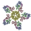
|
| 4 | x 6
|
| 5 |
|
| Symmetry | Point symmetry: (Schoenflies symbol: I (icosahedral)) |
-Components
| #1: Protein | Mass: 38428.023 Da / Num. of mol.: 4 Source method: isolated from a genetically manipulated source Source: (gene. exp.) Vibrio phage ICP1_2011_A (virus) / Gene: ICP12011A_121 / Plasmid: pETDUET / Production host: #2: Protein | | Mass: 33231.152 Da / Num. of mol.: 1 Source method: isolated from a genetically manipulated source Source: (gene. exp.) Vibrio cholerae (bacteria) / Strain: El Tor / Variant: PLE1 / Plasmid: pETDUET / Production host: Has protein modification | N | |
|---|
-Experimental details
-Experiment
| Experiment | Method: ELECTRON MICROSCOPY |
|---|---|
| EM experiment | Aggregation state: PARTICLE / 3D reconstruction method: single particle reconstruction |
- Sample preparation
| Component | Name: PLE Procapsid / Type: COMPLEX / Entity ID: all / Source: MULTIPLE SOURCES | |||||||||||||||||||||||||
|---|---|---|---|---|---|---|---|---|---|---|---|---|---|---|---|---|---|---|---|---|---|---|---|---|---|---|
| Molecular weight | Experimental value: NO | |||||||||||||||||||||||||
| Source (natural) | Organism: Vibrio cholerae (bacteria) / Strain: El Tor | |||||||||||||||||||||||||
| Source (recombinant) | Organism: | |||||||||||||||||||||||||
| Details of virus | Empty: NO / Enveloped: NO / Isolate: OTHER / Type: VIRION | |||||||||||||||||||||||||
| Natural host | Organism: Shigella flexneri Y | |||||||||||||||||||||||||
| Buffer solution | pH: 7.5 Details: 50 mM Tris-HCl, pH 7.4, 100 mM NaCl, 10 mM MgSO4, 1 mM CaCl2 | |||||||||||||||||||||||||
| Buffer component |
| |||||||||||||||||||||||||
| Specimen | Embedding applied: NO / Shadowing applied: NO / Staining applied: NO / Vitrification applied: YES | |||||||||||||||||||||||||
| Vitrification | Instrument: FEI VITROBOT MARK IV / Cryogen name: ETHANE / Humidity: 100 % / Chamber temperature: 277.15 K |
- Electron microscopy imaging
| Experimental equipment |  Model: Titan Krios / Image courtesy: FEI Company |
|---|---|
| Microscopy | Model: FEI TITAN KRIOS |
| Electron gun | Electron source:  FIELD EMISSION GUN / Accelerating voltage: 300 kV / Illumination mode: FLOOD BEAM FIELD EMISSION GUN / Accelerating voltage: 300 kV / Illumination mode: FLOOD BEAM |
| Electron lens | Mode: BRIGHT FIELD / Nominal defocus max: 2000 nm / Nominal defocus min: 700 nm |
| Image recording | Electron dose: 36.41 e/Å2 / Film or detector model: GATAN K3 BIOQUANTUM (6k x 4k) |
- Processing
| CTF correction | Type: NONE | ||||||||||||||||||||||||
|---|---|---|---|---|---|---|---|---|---|---|---|---|---|---|---|---|---|---|---|---|---|---|---|---|---|
| Symmetry | Point symmetry: I (icosahedral) | ||||||||||||||||||||||||
| 3D reconstruction | Resolution: 3.4 Å / Resolution method: FSC 0.143 CUT-OFF / Num. of particles: 379643 / Symmetry type: POINT | ||||||||||||||||||||||||
| Refine LS restraints |
|
