 Movie
Movie Controller

[English] 日本語
 Yorodumi
- PDB-8g1l: Lectin domain of Aap (Staphylococcus epidermidis Accumulation Ass... -
+ Open data

- Basic information
| Entry | Database: PDB / ID: 8g1l | |||||||||
|---|---|---|---|---|---|---|---|---|---|---|
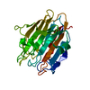
| Title | Lectin domain of Aap (Staphylococcus epidermidis Accumulation Associated Protein) | |||||||||
 Components Components | Accumulation associated protein | |||||||||
 Keywords Keywords | CELL ADHESION / bacterial lectin / cell wall-attached protein / glycan binding | |||||||||
| Function / homology |  Function and homology information Function and homology information | |||||||||
| Biological species |  Staphylococcus epidermidis RP62A (bacteria) Staphylococcus epidermidis RP62A (bacteria) | |||||||||
| Method |  X-RAY DIFFRACTION / SYNCHROTRON / SAD / Resolution: 1.06 Å X-RAY DIFFRACTION / SYNCHROTRON / SAD / Resolution: 1.06 Å | |||||||||
 Authors Authors | Maciag, J.J. / Herr, A.B. | |||||||||
| Funding support |  United States, 2items United States, 2items
| |||||||||
 Citation Citation | Journal: To Be Published Title: Mechanistic basis of staphylococcal interspecies competition for skin colonization Authors: Maciag, J.J. / Herr, A.B. | |||||||||
| History |
|
- Structure visualization
| Structure viewer | Molecule: MolmilJmol/JSmol |
|---|
- Downloads & links
-Download
| PDBx/mmCIF format | 8g1l.cif.gz | 169 KB | Display | PDBx/mmCIF format |
|---|---|---|---|---|
| PDB format | pdb8g1l.ent.gz | 133.8 KB | Display | PDB format |
| PDBx/mmJSON format | 8g1l.json.gz | Tree view | PDBx/mmJSON format | |
| Others |  Other downloads Other downloads |
-Validation report
| Summary document | 8g1l_validation.pdf.gz | 427.5 KB | Display | wwPDB validaton report |
|---|---|---|---|---|
| Full document | 8g1l_full_validation.pdf.gz | 427.9 KB | Display | |
| Data in XML | 8g1l_validation.xml.gz | 14.6 KB | Display | |
| Data in CIF | 8g1l_validation.cif.gz | 22.7 KB | Display | |
| Arichive directory | https://data.pdbj.org/pub/pdb/validation_reports/g1/8g1lftp://data.pdbj.org/pub/pdb/validation_reports/g1/8g1l | HTTPS FTP |
-Related structure data
-Links
 PDBj
PDBj- Assembly
| Deposited unit | 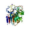
| ||||||||
|---|---|---|---|---|---|---|---|---|---|
| 1 |
| ||||||||
| Unit cell |
|
-Components
-Protein , 1 types, 1 molecules A
| #1: Protein | Mass: 27437.576 Da / Num. of mol.: 1 Source method: isolated from a genetically manipulated source Source: (gene. exp.) Staphylococcus epidermidis RP62A (bacteria)Gene: aap, SERP2398 / Plasmid: pDest-HisMBP / Production host: |
|---|
-Non-polymers , 5 types, 352 molecules 


| #2: Chemical | ChemComp-CA /  Mass: 40.078 Da / Num. of mol.: 1 / Source method: obtained synthetically / Formula: Ca Mass: 40.078 Da / Num. of mol.: 1 / Source method: obtained synthetically / Formula: Ca | ||||||
|---|---|---|---|---|---|---|---|
| #3: Chemical |  Mass: 24.305 Da / Num. of mol.: 3 / Source method: obtained synthetically / Formula: Mg Mass: 24.305 Da / Num. of mol.: 3 / Source method: obtained synthetically / Formula: Mg#4: Chemical |  Mass: 22.990 Da / Num. of mol.: 2 / Source method: obtained synthetically / Formula: Na Mass: 22.990 Da / Num. of mol.: 2 / Source method: obtained synthetically / Formula: Na#5: Chemical | ChemComp-PG0 / |  Mass: 120.147 Da / Num. of mol.: 1 / Source method: obtained synthetically / Formula: C5H12O3 / Comment: precipitant*YM Mass: 120.147 Da / Num. of mol.: 1 / Source method: obtained synthetically / Formula: C5H12O3 / Comment: precipitant*YM#6: Water | ChemComp-HOH / | Mass: 18.015 Da / Num. of mol.: 345 / Source method: isolated from a natural source / Formula: H2O |
-Details
| Has ligand of interest | N |
|---|
-Experimental details
-Experiment
| Experiment | Method: X-RAY DIFFRACTION / Number of used crystals: 1 |
|---|
- Sample preparation
| Crystal | Density Matthews: 2.5 Å3/Da / Density % sol: 50.72 % |
|---|---|
| Crystal grow | Temperature: 295 K / Method: vapor diffusion, hanging drop Details: 100 mM Na Acetate (pH 4.5), 22.5% BCS PEG SMEAR High (Molecular Dimensions, CalibreScientific), pentanediol and 100 mM BisTris (pH 6.5) |
-Data collection
| Diffraction | Mean temperature: 183 K / Serial crystal experiment: N |
|---|---|
| Diffraction source | Source: SYNCHROTRON / Site: APS / Beamline: 24-ID-C / Wavelength: 0.9778 Å |
| Detector | Type: DECTRIS EIGER2 X 16M / Detector: PIXEL / Date: Nov 17, 2018 |
| Radiation | Protocol: SINGLE WAVELENGTH / Monochromatic (M) / Laue (L): M / Scattering type: x-ray |
| Radiation wavelength | Wavelength: 0.9778 Å / Relative weight: 1 |
| Reflection | Resolution: 1.06→42.7658 Å / Num. obs: 121879 / % possible obs: 99.65 % / Redundancy: 8.2 % / CC1/2: 0.997 / Rmerge(I) obs: 0.09085 / Rpim(I) all: 0.03388 / Rrim(I) all: 0.09719 / Net I/σ(I): 14.88 |
| Reflection shell | Resolution: 1.06→1.098 Å / Redundancy: 7.5 % / Rmerge(I) obs: 0.5542 / Mean I/σ(I) obs: 4.39 / Num. unique obs: 11899 / CC1/2: 0.911 / Rpim(I) all: 0.2158 / Rrim(I) all: 0.596 / % possible all: 98.48 |
- Processing
| Software |
| |||||||||||||||||||||||||||||||||||||||||||||||||||||||||||||||||||||||||||||||||||||||||||||||||||||||||
|---|---|---|---|---|---|---|---|---|---|---|---|---|---|---|---|---|---|---|---|---|---|---|---|---|---|---|---|---|---|---|---|---|---|---|---|---|---|---|---|---|---|---|---|---|---|---|---|---|---|---|---|---|---|---|---|---|---|---|---|---|---|---|---|---|---|---|---|---|---|---|---|---|---|---|---|---|---|---|---|---|---|---|---|---|---|---|---|---|---|---|---|---|---|---|---|---|---|---|---|---|---|---|---|---|---|---|
| Refinement | Method to determine structure: SAD / Resolution: 1.06→42.7658 Å / SU ML: 0.05 / Cross valid method: FREE R-VALUE / σ(F): 1.37 / Phase error: 9.61 / Stereochemistry target values: ML
| |||||||||||||||||||||||||||||||||||||||||||||||||||||||||||||||||||||||||||||||||||||||||||||||||||||||||
| Solvent computation | Shrinkage radii: 0.9 Å / VDW probe radii: 1.11 Å / Solvent model: FLAT BULK SOLVENT MODEL | |||||||||||||||||||||||||||||||||||||||||||||||||||||||||||||||||||||||||||||||||||||||||||||||||||||||||
| Refinement step | Cycle: LAST / Resolution: 1.06→42.7658 Å
| |||||||||||||||||||||||||||||||||||||||||||||||||||||||||||||||||||||||||||||||||||||||||||||||||||||||||
| Refine LS restraints |
| |||||||||||||||||||||||||||||||||||||||||||||||||||||||||||||||||||||||||||||||||||||||||||||||||||||||||
| LS refinement shell |
|
