 Movie
Movie Controller

[English] 日本語
 Yorodumi
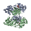
- PDB-8g0f: Crystal structure of diphtheria toxin H223Q/H257Q double mutant (... -
+ Open data

- Basic information
| Entry | Database: PDB / ID: 8g0f | ||||||
|---|---|---|---|---|---|---|---|
| Title | Crystal structure of diphtheria toxin H223Q/H257Q double mutant (pH 5.5) | ||||||
 Components Components | Diphtheria toxin | ||||||
 Keywords Keywords | TOXIN / diptheria toxin / pH dependent conformational changes | ||||||
| Function / homology |  Function and homology information Function and homology information | ||||||
| Biological species |  Corynebacterium diphtheriae (bacteria) Corynebacterium diphtheriae (bacteria) | ||||||
| Method |  X-RAY DIFFRACTION / SYNCHROTRON / MOLECULAR REPLACEMENT / Resolution: 2.25 Å X-RAY DIFFRACTION / SYNCHROTRON / MOLECULAR REPLACEMENT / Resolution: 2.25 Å | ||||||
 Authors Authors | Lovell, S. / Kashipathy, M.M. / Battaile, K.P. / Ladokhin, A.S. | ||||||
| Funding support |  United States, 1items United States, 1items
| ||||||
 Citation Citation | Journal: Toxins / Year: 2023 Title: Histidine Protonation and Conformational Switching in Diphtheria Toxin Translocation Domain. Authors: Rodnin, M.V. / Vasques-Montes, V. / Kyrychenko, A. / Oliveira, N.F.B. / Kashipathy, M.M. / Battaile, K.P. / Douglas, J. / Lovell, S. / Machuqueiro, M. / Ladokhin, A.S. | ||||||
| History |
|
- Structure visualization
| Structure viewer | Molecule: MolmilJmol/JSmol |
|---|
- Downloads & links
-Download
| PDBx/mmCIF format | 8g0f.cif.gz | 204.4 KB | Display | PDBx/mmCIF format |
|---|---|---|---|---|
| PDB format | pdb8g0f.ent.gz | 158.8 KB | Display | PDB format |
| PDBx/mmJSON format | 8g0f.json.gz | Tree view | PDBx/mmJSON format | |
| Others |  Other downloads Other downloads |
-Validation report
| Arichive directory | https://data.pdbj.org/pub/pdb/validation_reports/g0/8g0fftp://data.pdbj.org/pub/pdb/validation_reports/g0/8g0f | HTTPS FTP |
|---|
-Related structure data
-Links
 PDBj
PDBj
- Assembly
| Deposited unit | 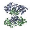
| ||||||||
|---|---|---|---|---|---|---|---|---|---|
| 1 |
| ||||||||
| Unit cell |
|
-Components
| #1: Protein | Mass: 58724.797 Da / Num. of mol.: 2 / Mutation: K51E, E148K, H223Q, H257Q Source method: isolated from a genetically manipulated source Source: (gene. exp.) Corynebacterium diphtheriae (bacteria) / Plasmid: pET15b / Production host: #2: Water | ChemComp-HOH / |  Mass: 18.015 Da / Num. of mol.: 167 / Source method: isolated from a natural source / Formula: H2O Mass: 18.015 Da / Num. of mol.: 167 / Source method: isolated from a natural source / Formula: H2OHas protein modification | Y | |
|---|
-Experimental details
-Experiment
| Experiment | Method: X-RAY DIFFRACTION / Number of used crystals: 1 |
|---|
- Sample preparation
| Crystal | Density Matthews: 2.58 Å3/Da / Density % sol: 52.26 % |
|---|---|
| Crystal grow | Temperature: 291 K / Method: vapor diffusion, sitting drop / pH: 5.5 Details: 20% (w/v) PEG 2000 MME, 100 mM sodium citrate tribasic, 200 mM sodium malonate |
-Data collection
| Diffraction | Mean temperature: 100 K / Serial crystal experiment: N |
|---|---|
| Diffraction source | Source: SYNCHROTRON / Site: APS / Beamline: 17-ID / Wavelength: 1 Å |
| Detector | Type: DECTRIS PILATUS 6M / Detector: PIXEL / Date: Jun 23, 2018 |
| Radiation | Protocol: SINGLE WAVELENGTH / Monochromatic (M) / Laue (L): M / Scattering type: x-ray |
| Radiation wavelength | Wavelength: 1 Å / Relative weight: 1 |
| Reflection | Resolution: 2.25→46.78 Å / Num. obs: 52984 / % possible obs: 95.8 % / Redundancy: 3.4 % / CC1/2: 0.997 / Rmerge(I) obs: 0.074 / Rpim(I) all: 0.047 / Rrim(I) all: 0.088 / Χ2: 0.92 / Net I/σ(I): 9.7 / Num. measured all: 182683 |
| Reflection shell | Resolution: 2.25→2.32 Å / % possible obs: 96.7 % / Redundancy: 3.4 % / Rmerge(I) obs: 0.727 / Num. measured all: 15664 / Num. unique obs: 4660 / CC1/2: 0.692 / Rpim(I) all: 0.469 / Rrim(I) all: 0.869 / Χ2: 0.81 / Net I/σ(I) obs: 1.7 |
- Processing
| Software |
| ||||||||||||||||||||||||||||||||||||||||||||||||||||||||||||||||||||||||||||||||||||||||||||||||||||||||||||||||||||||||||||||||||||||||||||
|---|---|---|---|---|---|---|---|---|---|---|---|---|---|---|---|---|---|---|---|---|---|---|---|---|---|---|---|---|---|---|---|---|---|---|---|---|---|---|---|---|---|---|---|---|---|---|---|---|---|---|---|---|---|---|---|---|---|---|---|---|---|---|---|---|---|---|---|---|---|---|---|---|---|---|---|---|---|---|---|---|---|---|---|---|---|---|---|---|---|---|---|---|---|---|---|---|---|---|---|---|---|---|---|---|---|---|---|---|---|---|---|---|---|---|---|---|---|---|---|---|---|---|---|---|---|---|---|---|---|---|---|---|---|---|---|---|---|---|---|---|---|
| Refinement | Method to determine structure: MOLECULAR REPLACEMENT / Resolution: 2.25→36.31 Å / SU ML: 0.33 / Cross valid method: FREE R-VALUE / σ(F): 1.15 / Phase error: 30.18 / Stereochemistry target values: ML
| ||||||||||||||||||||||||||||||||||||||||||||||||||||||||||||||||||||||||||||||||||||||||||||||||||||||||||||||||||||||||||||||||||||||||||||
| Solvent computation | Shrinkage radii: 0.9 Å / VDW probe radii: 1.11 Å / Solvent model: FLAT BULK SOLVENT MODEL | ||||||||||||||||||||||||||||||||||||||||||||||||||||||||||||||||||||||||||||||||||||||||||||||||||||||||||||||||||||||||||||||||||||||||||||
| Refinement step | Cycle: LAST / Resolution: 2.25→36.31 Å
| ||||||||||||||||||||||||||||||||||||||||||||||||||||||||||||||||||||||||||||||||||||||||||||||||||||||||||||||||||||||||||||||||||||||||||||
| Refine LS restraints |
| ||||||||||||||||||||||||||||||||||||||||||||||||||||||||||||||||||||||||||||||||||||||||||||||||||||||||||||||||||||||||||||||||||||||||||||
| LS refinement shell |
|
