 Movie
Movie Controller


+ Open data

- Basic information
| Entry | Database: PDB / ID: 8fzb | ||||||
|---|---|---|---|---|---|---|---|
| Title | Crystal structure of human FAM86A | ||||||
 Components Components | Protein-lysine N-methyltransferase EEF2KMT | ||||||
 Keywords Keywords | TRANSFERASE / Protein Lysine methyltransferase | ||||||
| Function / homology |  Function and homology information Function and homology informationpeptidyl-lysine trimethylation / protein-lysine N-methyltransferase activity / histone methyltransferase activity / Protein methylation / Transferases; Transferring one-carbon groups; Methyltransferases / protein-containing complex / cytosol / cytoplasm Similarity search - Function | ||||||
| Biological species |  Homo sapiens (human) Homo sapiens (human) | ||||||
| Method |  X-RAY DIFFRACTION / SYNCHROTRON / MOLECULAR REPLACEMENT / Resolution: 3.35 Å X-RAY DIFFRACTION / SYNCHROTRON / MOLECULAR REPLACEMENT / Resolution: 3.35 Å | ||||||
 Authors Authors | Shao, Z. / Lu, J. / Song, J. | ||||||
| Funding support |  United States, 1items United States, 1items
| ||||||
 Citation Citation | Journal: J.Biol.Chem. / Year: 2023 Title: The FAM86 domain of FAM86A confers substrate specificity to promote EEF2-Lys525 methylation. Authors: Francis, J.W. / Shao, Z. / Narkhede, P. / Trinh, A.T. / Lu, J. / Song, J. / Gozani, O. | ||||||
| History |
|
- Structure visualization
| Structure viewer | Molecule: MolmilJmol/JSmol |
|---|
- Downloads & links
-Download
| PDBx/mmCIF format | 8fzb.cif.gz | 193.9 KB | Display | PDBx/mmCIF format |
|---|---|---|---|---|
| PDB format | pdb8fzb.ent.gz | 153.9 KB | Display | PDB format |
| PDBx/mmJSON format | 8fzb.json.gz | Tree view | PDBx/mmJSON format | |
| Others |  Other downloads Other downloads |
-Validation report
| Summary document | 8fzb_validation.pdf.gz | 1.2 MB | Display | wwPDB validaton report |
|---|---|---|---|---|
| Full document | 8fzb_full_validation.pdf.gz | 1.2 MB | Display | |
| Data in XML | 8fzb_validation.xml.gz | 34.3 KB | Display | |
| Data in CIF | 8fzb_validation.cif.gz | 45.4 KB | Display | |
| Arichive directory | https://data.pdbj.org/pub/pdb/validation_reports/fz/8fzbftp://data.pdbj.org/pub/pdb/validation_reports/fz/8fzb | HTTPS FTP |
-Related structure data
| Similar structure data |
|---|
-Links
 PDBj
PDBj


- Assembly
| Deposited unit | 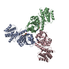
| ||||||||
|---|---|---|---|---|---|---|---|---|---|
| 1 | 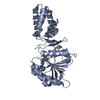
| ||||||||
| 2 | 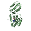
| ||||||||
| 3 | 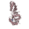
| ||||||||
| Unit cell |
|
-Components
| #1: Protein | Mass: 37132.445 Da / Num. of mol.: 3 Source method: isolated from a genetically manipulated source Source: (gene. exp.) Homo sapiens (human) / Gene: EEF2KMT, FAM86A, SB153 / Production host:  References: UniProt: Q96G04, Transferases; Transferring one-carbon groups; Methyltransferases #2: Chemical |   Type: L-peptide linking / Mass: 384.411 Da / Num. of mol.: 3 / Source method: obtained synthetically / Formula: C14H20N6O5S / Feature type: SUBJECT OF INVESTIGATION Type: L-peptide linking / Mass: 384.411 Da / Num. of mol.: 3 / Source method: obtained synthetically / Formula: C14H20N6O5S / Feature type: SUBJECT OF INVESTIGATIONHas ligand of interest | Y | |
|---|
-Experimental details
-Experiment
| Experiment | Method: X-RAY DIFFRACTION / Number of used crystals: 1 |
|---|
- Sample preparation
| Crystal | Density Matthews: 5.1 Å3/Da / Density % sol: 75.87 % |
|---|---|
| Crystal grow | Temperature: 285 K / Method: vapor diffusion, hanging drop Details: 0.1 M Ammonium citrate tribasic (pH 7.0), 10% w/v Polyethylene glycol 3,350 and 5 mM TCEP |
-Data collection
| Diffraction | Mean temperature: 100 K / Serial crystal experiment: N | |||||||||||||||||||||||||||||||||||||||||||||||||||||||||||||||||||||||||||||
|---|---|---|---|---|---|---|---|---|---|---|---|---|---|---|---|---|---|---|---|---|---|---|---|---|---|---|---|---|---|---|---|---|---|---|---|---|---|---|---|---|---|---|---|---|---|---|---|---|---|---|---|---|---|---|---|---|---|---|---|---|---|---|---|---|---|---|---|---|---|---|---|---|---|---|---|---|---|---|
| Diffraction source | Source: SYNCHROTRON / Site: APS / Beamline: 24-ID-C / Wavelength: 0.97918 Å | |||||||||||||||||||||||||||||||||||||||||||||||||||||||||||||||||||||||||||||
| Detector | Type: DECTRIS EIGER2 X 16M / Detector: PIXEL / Date: Apr 10, 2022 | |||||||||||||||||||||||||||||||||||||||||||||||||||||||||||||||||||||||||||||
| Radiation | Protocol: SINGLE WAVELENGTH / Monochromatic (M) / Laue (L): M / Scattering type: x-ray | |||||||||||||||||||||||||||||||||||||||||||||||||||||||||||||||||||||||||||||
| Radiation wavelength | Wavelength: 0.97918 Å / Relative weight: 1 | |||||||||||||||||||||||||||||||||||||||||||||||||||||||||||||||||||||||||||||
| Reflection | Resolution: 3.35→50 Å / Num. obs: 64154 / % possible obs: 100 % / Redundancy: 20.7 % / Rmerge(I) obs: 0.998 / Χ2: 0.033 / Net I/σ(I): 4.5 / Num. measured all: 1324884 | |||||||||||||||||||||||||||||||||||||||||||||||||||||||||||||||||||||||||||||
| Reflection shell |
|
- Processing
| Software |
| |||||||||||||||||||||||||||||||||||||||||||||||||||||||||||||||||||||||||||||||||||||||||||||||||||||||||
|---|---|---|---|---|---|---|---|---|---|---|---|---|---|---|---|---|---|---|---|---|---|---|---|---|---|---|---|---|---|---|---|---|---|---|---|---|---|---|---|---|---|---|---|---|---|---|---|---|---|---|---|---|---|---|---|---|---|---|---|---|---|---|---|---|---|---|---|---|---|---|---|---|---|---|---|---|---|---|---|---|---|---|---|---|---|---|---|---|---|---|---|---|---|---|---|---|---|---|---|---|---|---|---|---|---|---|
| Refinement | Method to determine structure: MOLECULAR REPLACEMENT / Resolution: 3.35→48.82 Å / SU ML: 0.55 / Cross valid method: FREE R-VALUE / σ(F): 1.34 / Phase error: 29.17 / Stereochemistry target values: ML
| |||||||||||||||||||||||||||||||||||||||||||||||||||||||||||||||||||||||||||||||||||||||||||||||||||||||||
| Solvent computation | Shrinkage radii: 0.9 Å / VDW probe radii: 1.1 Å / Solvent model: FLAT BULK SOLVENT MODEL | |||||||||||||||||||||||||||||||||||||||||||||||||||||||||||||||||||||||||||||||||||||||||||||||||||||||||
| Refinement step | Cycle: LAST / Resolution: 3.35→48.82 Å
| |||||||||||||||||||||||||||||||||||||||||||||||||||||||||||||||||||||||||||||||||||||||||||||||||||||||||
| Refine LS restraints |
| |||||||||||||||||||||||||||||||||||||||||||||||||||||||||||||||||||||||||||||||||||||||||||||||||||||||||
| LS refinement shell |
|
