 Movie
Movie Controller

[English] 日本語
 Yorodumi
- PDB-8fxt: Escherichia coli periplasmic Glucose-Binding Protein glucose comp... -
+ Open data

- Basic information
| Entry | Database: PDB / ID: 8fxt | ||||||
|---|---|---|---|---|---|---|---|
| Title | Escherichia coli periplasmic Glucose-Binding Protein glucose complex: Acrylodan conjugate attached at W183C | ||||||
 Components Components | D-galactose/methyl-galactoside binding periplasmic protein MglB | ||||||
 Keywords Keywords | SUGAR BINDING PROTEIN / Periplasmic binding protein / Biosensor Fluorescent conjugate | ||||||
| Function / homology |  Function and homology information Function and homology informationouter membrane-bounded periplasmic space / carbohydrate binding / metal ion binding Similarity search - Function | ||||||
| Biological species |  | ||||||
| Method |  X-RAY DIFFRACTION / SYNCHROTRON / MOLECULAR REPLACEMENT / Resolution: 1.53 Å X-RAY DIFFRACTION / SYNCHROTRON / MOLECULAR REPLACEMENT / Resolution: 1.53 Å | ||||||
 Authors Authors | Allert, M.J. / Kumar, S. / Wang, Y. / Beese, L.S. / Hellinga, H.W. | ||||||
| Funding support |  United States, 1items United States, 1items
| ||||||
 Citation Citation | Journal: Commun Chem / Year: 2023 Title: Chromophore carbonyl twisting in fluorescent biosensors encodes direct readout of protein conformations with multicolor switching. Authors: Allert, M.J. / Kumar, S. / Wang, Y. / Beese, L.S. / Hellinga, H.W. | ||||||
| History |
|
- Structure visualization
| Structure viewer | Molecule: MolmilJmol/JSmol |
|---|
- Downloads & links
-Download
| PDBx/mmCIF format | 8fxt.cif.gz | 91.5 KB | Display | PDBx/mmCIF format |
|---|---|---|---|---|
| PDB format | pdb8fxt.ent.gz | 61.3 KB | Display | PDB format |
| PDBx/mmJSON format | 8fxt.json.gz | Tree view | PDBx/mmJSON format | |
| Others |  Other downloads Other downloads |
-Validation report
| Summary document | 8fxt_validation.pdf.gz | 689.1 KB | Display | wwPDB validaton report |
|---|---|---|---|---|
| Full document | 8fxt_full_validation.pdf.gz | 689.2 KB | Display | |
| Data in XML | 8fxt_validation.xml.gz | 16.4 KB | Display | |
| Data in CIF | 8fxt_validation.cif.gz | 24.9 KB | Display | |
| Arichive directory | https://data.pdbj.org/pub/pdb/validation_reports/fx/8fxtftp://data.pdbj.org/pub/pdb/validation_reports/fx/8fxt | HTTPS FTP |
-Related structure data
| Related structure data | 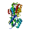 8fxuC C: citing same article ( |
|---|---|
| Similar structure data | |
| Experimental dataset #1 | Data reference: 10.5061/dryad.msbcc2g2x / Data set type: other data |
-Links
 PDBj
PDBj


- Assembly
| Deposited unit | 
| ||||||||||
|---|---|---|---|---|---|---|---|---|---|---|---|
| 1 |
| ||||||||||
| Unit cell |
| ||||||||||
| Components on special symmetry positions |
|
-Components
-Protein , 1 types, 1 molecules A
| #1: Protein | Mass: 32864.027 Da / Num. of mol.: 1 / Mutation: W183C Source method: isolated from a genetically manipulated source Source: (gene. exp.) |
|---|
-Sugars , 2 types, 6 molecules 

| #3: Sugar | ChemComp-GLC /  Type: D-saccharide, alpha linking / Mass: 180.156 Da / Num. of mol.: 1 / Source method: obtained synthetically / Formula: C6H12O6 Type: D-saccharide, alpha linking / Mass: 180.156 Da / Num. of mol.: 1 / Source method: obtained synthetically / Formula: C6H12O6 |
|---|---|
| #4: Sugar | ChemComp-BGC /  Type: D-saccharide, beta linking / Mass: 180.156 Da / Num. of mol.: 5 / Source method: obtained synthetically / Formula: C6H12O6 Type: D-saccharide, beta linking / Mass: 180.156 Da / Num. of mol.: 5 / Source method: obtained synthetically / Formula: C6H12O6 |
-Non-polymers , 3 types, 320 molecules 


| #2: Chemical | ChemComp-YDM /  Mass: 227.302 Da / Num. of mol.: 1 / Source method: obtained synthetically / Formula: C15H17NO / Feature type: SUBJECT OF INVESTIGATION Mass: 227.302 Da / Num. of mol.: 1 / Source method: obtained synthetically / Formula: C15H17NO / Feature type: SUBJECT OF INVESTIGATION |
|---|---|
| #5: Chemical | ChemComp-CA /  Mass: 40.078 Da / Num. of mol.: 1 / Source method: obtained synthetically / Formula: Ca Mass: 40.078 Da / Num. of mol.: 1 / Source method: obtained synthetically / Formula: Ca |
| #6: Water | ChemComp-HOH / Mass: 18.015 Da / Num. of mol.: 318 / Source method: isolated from a natural source / Formula: H2O |
-Details
| Has ligand of interest | Y |
|---|---|
| Has protein modification | Y |
-Experimental details
-Experiment
| Experiment | Method: X-RAY DIFFRACTION / Number of used crystals: 1 |
|---|
- Sample preparation
| Crystal | Density Matthews: 2.19 Å3/Da / Density % sol: 43.9 % |
|---|---|
| Crystal grow | Temperature: 290 K / Method: vapor diffusion, hanging drop / Details: 20% PEG 3350 0.2M potassium thiocyanate |
-Data collection
| Diffraction | Mean temperature: 100 K / Serial crystal experiment: N |
|---|---|
| Diffraction source | Source: SYNCHROTRON / Site: APS / Beamline: 22-BM / Wavelength: 1 Å |
| Detector | Type: MARMOSAIC 225 mm CCD / Detector: CCD / Date: Nov 11, 2011 |
| Radiation | Protocol: SINGLE WAVELENGTH / Monochromatic (M) / Laue (L): M / Scattering type: x-ray |
| Radiation wavelength | Wavelength: 1 Å / Relative weight: 1 |
| Reflection | Resolution: 1.53→50 Å / Num. obs: 43030 / % possible obs: 98.8 % / Redundancy: 6.5 % / Biso Wilson estimate: 12.57 Å2 / Rmerge(I) obs: 0.06 / Rpim(I) all: 0.025 / Net I/σ(I): 25.54 |
| Reflection shell | Resolution: 1.53→1.56 Å / Rmerge(I) obs: 0.292 / Mean I/σ(I) obs: 3.99 / Num. unique obs: 2019 / Rpim(I) all: 0.131 |
- Processing
| Software |
| |||||||||||||||||||||||||||||||||||||||||||||||||||||||||||||||||||||||||||||||||||||||||||||||||||||||||
|---|---|---|---|---|---|---|---|---|---|---|---|---|---|---|---|---|---|---|---|---|---|---|---|---|---|---|---|---|---|---|---|---|---|---|---|---|---|---|---|---|---|---|---|---|---|---|---|---|---|---|---|---|---|---|---|---|---|---|---|---|---|---|---|---|---|---|---|---|---|---|---|---|---|---|---|---|---|---|---|---|---|---|---|---|---|---|---|---|---|---|---|---|---|---|---|---|---|---|---|---|---|---|---|---|---|---|
| Refinement | Method to determine structure: MOLECULAR REPLACEMENT / Resolution: 1.53→24.75 Å / SU ML: 0.149 / Cross valid method: FREE R-VALUE / σ(F): 1.38 / Phase error: 18.3363 Stereochemistry target values: GeoStd + Monomer Library + CDL v1.2
| |||||||||||||||||||||||||||||||||||||||||||||||||||||||||||||||||||||||||||||||||||||||||||||||||||||||||
| Solvent computation | Shrinkage radii: 0.9 Å / VDW probe radii: 1.1 Å / Solvent model: FLAT BULK SOLVENT MODEL | |||||||||||||||||||||||||||||||||||||||||||||||||||||||||||||||||||||||||||||||||||||||||||||||||||||||||
| Displacement parameters | Biso mean: 17.98 Å2 | |||||||||||||||||||||||||||||||||||||||||||||||||||||||||||||||||||||||||||||||||||||||||||||||||||||||||
| Refinement step | Cycle: LAST / Resolution: 1.53→24.75 Å
| |||||||||||||||||||||||||||||||||||||||||||||||||||||||||||||||||||||||||||||||||||||||||||||||||||||||||
| Refine LS restraints |
| |||||||||||||||||||||||||||||||||||||||||||||||||||||||||||||||||||||||||||||||||||||||||||||||||||||||||
| LS refinement shell |
|
