 Movie
Movie Controller


+ Open data

- Basic information
| Entry | Database: PDB / ID: 8fxd | |||||||||
|---|---|---|---|---|---|---|---|---|---|---|
| Title | Rubrerythrin from B. pseudomallei: manganese-bound | |||||||||
 Components Components | Rubrerythrin | |||||||||
 Keywords Keywords | OXIDOREDUCTASE / metalloprotein / oxidative stress / non-heme | |||||||||
| Function / homology |  Function and homology information Function and homology information | |||||||||
| Biological species |  Burkholderia pseudomallei (bacteria) Burkholderia pseudomallei (bacteria) | |||||||||
| Method |  X-RAY DIFFRACTION / SYNCHROTRON / MOLECULAR REPLACEMENT / Resolution: 1.58 Å X-RAY DIFFRACTION / SYNCHROTRON / MOLECULAR REPLACEMENT / Resolution: 1.58 Å | |||||||||
 Authors Authors | Monteiro, D.C.F. / Snell, M.E. / Budziszewski, G.R. / Bowman, S.E.J. | |||||||||
| Funding support |  United States, 2items United States, 2items
| |||||||||
 Citation Citation | Journal: Biorxiv / Year: 2025 Title: Burkholderia pseudomallei rubrerythrin promiscuously binds metals in a structurally pre-formed bimetallic binding site. Authors: Budziszewski, G.R. / Lynch, M.L. / Snell, M.E. / Monteiro, D.C. / Bowman, S.E. | |||||||||
| History |
|
- Structure visualization
| Structure viewer | Molecule: MolmilJmol/JSmol |
|---|
- Downloads & links
-Download
| PDBx/mmCIF format | 8fxd.cif.gz | 343.7 KB | Display | PDBx/mmCIF format |
|---|---|---|---|---|
| PDB format | pdb8fxd.ent.gz | 270.9 KB | Display | PDB format |
| PDBx/mmJSON format | 8fxd.json.gz | Tree view | PDBx/mmJSON format | |
| Others |  Other downloads Other downloads |
-Validation report
| Summary document | 8fxd_validation.pdf.gz | 493.9 KB | Display | wwPDB validaton report |
|---|---|---|---|---|
| Full document | 8fxd_full_validation.pdf.gz | 497.3 KB | Display | |
| Data in XML | 8fxd_validation.xml.gz | 41.1 KB | Display | |
| Data in CIF | 8fxd_validation.cif.gz | 60.7 KB | Display | |
| Arichive directory | https://data.pdbj.org/pub/pdb/validation_reports/fx/8fxdftp://data.pdbj.org/pub/pdb/validation_reports/fx/8fxd | HTTPS FTP |
-Related structure data
| Related structure data |  8fuhC 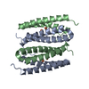 8fvvC  9onmC  9onnC  9onoC  9onqC  9onrC C: citing same article ( |
|---|---|
| Similar structure data |
-Links
 PDBj
PDBj


- Assembly
| Deposited unit | 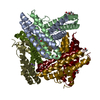
| ||||||||
|---|---|---|---|---|---|---|---|---|---|
| 1 | 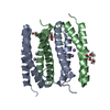
| ||||||||
| 2 | 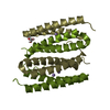
| ||||||||
| 3 | 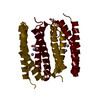
| ||||||||
| Unit cell |
|
-Components
| #1: Protein | Mass: 18655.389 Da / Num. of mol.: 6 Source method: isolated from a genetically manipulated source Source: (gene. exp.) Burkholderia pseudomallei (bacteria) / Gene: BURPS1710b_A0924 / Production host: #2: Chemical | ChemComp-PEG /   Mass: 106.120 Da / Num. of mol.: 7 / Source method: obtained synthetically / Formula: C4H10O3 Mass: 106.120 Da / Num. of mol.: 7 / Source method: obtained synthetically / Formula: C4H10O3#3: Chemical | ChemComp-MN /   Mass: 54.938 Da / Num. of mol.: 12 / Source method: obtained synthetically / Formula: Mn Mass: 54.938 Da / Num. of mol.: 12 / Source method: obtained synthetically / Formula: Mn#4: Water | ChemComp-HOH / |  Mass: 18.015 Da / Num. of mol.: 874 / Source method: isolated from a natural source / Formula: H2O Mass: 18.015 Da / Num. of mol.: 874 / Source method: isolated from a natural source / Formula: H2OHas ligand of interest | N | Has protein modification | N | |
|---|
-Experimental details
-Experiment
| Experiment | Method: X-RAY DIFFRACTION / Number of used crystals: 1 |
|---|
- Sample preparation
| Crystal | Density Matthews: 2.42 Å3/Da / Density % sol: 49.14 % |
|---|---|
| Crystal grow | Temperature: 295 K / Method: vapor diffusion, sitting drop / pH: 8 Details: 0.138 M Li2SO4, 0.92 M BTP, pH 8, 22.08% PEG 3350, 0.8 mM MnCl2 |
-Data collection
| Diffraction | Mean temperature: 100 K / Serial crystal experiment: N |
|---|---|
| Diffraction source | Source: SYNCHROTRON / Site: NSLS-II / Beamline: 17-ID-2 / Wavelength: 0.97934 Å |
| Detector | Type: DECTRIS EIGER X 16M / Detector: PIXEL / Date: Jun 22, 2022 |
| Radiation | Protocol: SINGLE WAVELENGTH / Monochromatic (M) / Laue (L): M / Scattering type: x-ray |
| Radiation wavelength | Wavelength: 0.97934 Å / Relative weight: 1 |
| Reflection | Resolution: 1.58→29.14 Å / Num. obs: 108953 / % possible obs: 93.7 % / Redundancy: 5.2 % / CC1/2: 0.998 / Rmerge(I) obs: 0.092 / Rpim(I) all: 0.069 / Rrim(I) all: 0.115 / Net I/σ(I): 9.5 |
| Reflection shell | Resolution: 1.58→1.692 Å / Redundancy: 3.1 % / Rmerge(I) obs: 0.833 / Num. unique obs: 5450 / CC1/2: 0.457 / Rpim(I) all: 0.807 / Rrim(I) all: 1.161 / % possible all: 59.3 |
- Processing
| Software |
| ||||||||||||||||||||||||||||||||||||||||||||||||||||||||||||||||||||||||||||||||||||||||||||||||||||||||||||||||||||||||||||||||||||||||||||||||||||||
|---|---|---|---|---|---|---|---|---|---|---|---|---|---|---|---|---|---|---|---|---|---|---|---|---|---|---|---|---|---|---|---|---|---|---|---|---|---|---|---|---|---|---|---|---|---|---|---|---|---|---|---|---|---|---|---|---|---|---|---|---|---|---|---|---|---|---|---|---|---|---|---|---|---|---|---|---|---|---|---|---|---|---|---|---|---|---|---|---|---|---|---|---|---|---|---|---|---|---|---|---|---|---|---|---|---|---|---|---|---|---|---|---|---|---|---|---|---|---|---|---|---|---|---|---|---|---|---|---|---|---|---|---|---|---|---|---|---|---|---|---|---|---|---|---|---|---|---|---|---|---|---|
| Refinement | Method to determine structure: MOLECULAR REPLACEMENT / Resolution: 1.58→29.14 Å / Cor.coef. Fo:Fc: 0.973 / Cor.coef. Fo:Fc free: 0.962 / SU B: 1.824 / SU ML: 0.059 / Cross valid method: FREE R-VALUE / ESU R: 0.083 / ESU R Free: 0.083 Details: Hydrogens have been added in their riding positions
| ||||||||||||||||||||||||||||||||||||||||||||||||||||||||||||||||||||||||||||||||||||||||||||||||||||||||||||||||||||||||||||||||||||||||||||||||||||||
| Solvent computation | Ion probe radii: 0.8 Å / Shrinkage radii: 0.8 Å / VDW probe radii: 1.2 Å / Solvent model: MASK BULK SOLVENT | ||||||||||||||||||||||||||||||||||||||||||||||||||||||||||||||||||||||||||||||||||||||||||||||||||||||||||||||||||||||||||||||||||||||||||||||||||||||
| Displacement parameters | Biso mean: 19.178 Å2
| ||||||||||||||||||||||||||||||||||||||||||||||||||||||||||||||||||||||||||||||||||||||||||||||||||||||||||||||||||||||||||||||||||||||||||||||||||||||
| Refinement step | Cycle: LAST / Resolution: 1.58→29.14 Å
| ||||||||||||||||||||||||||||||||||||||||||||||||||||||||||||||||||||||||||||||||||||||||||||||||||||||||||||||||||||||||||||||||||||||||||||||||||||||
| Refine LS restraints |
| ||||||||||||||||||||||||||||||||||||||||||||||||||||||||||||||||||||||||||||||||||||||||||||||||||||||||||||||||||||||||||||||||||||||||||||||||||||||
| LS refinement shell |
|
