 Movie
Movie Controller


+ Open data

- Basic information
| Entry | Database: PDB / ID: 8fw1 | |||||||||
|---|---|---|---|---|---|---|---|---|---|---|
| Title | Gluconobacter Ene-Reductase (GluER) mutant - PagER | |||||||||
 Components Components | N-ethylmaleimide reductase | |||||||||
 Keywords Keywords | OXIDOREDUCTASE / flavoprotein / 'ene'-reductase / photoenzyme | |||||||||
| Function / homology | oxidoreductase activity, acting on the CH-CH group of donors, NAD or NADP as acceptor / Oxidoreductase Oye-like / NADH:flavin oxidoreductase/NADH oxidase, N-terminal / NADH:flavin oxidoreductase / NADH oxidase family / FMN binding / Aldolase-type TIM barrel / cytosol / FLAVIN MONONUCLEOTIDE / Alkene reductase Function and homology information Function and homology information | |||||||||
| Biological species |  Gluconobacter oxydans (bacteria) Gluconobacter oxydans (bacteria) | |||||||||
| Method |  X-RAY DIFFRACTION / SYNCHROTRON / MOLECULAR REPLACEMENT / Resolution: 1.5 Å X-RAY DIFFRACTION / SYNCHROTRON / MOLECULAR REPLACEMENT / Resolution: 1.5 Å | |||||||||
 Authors Authors | Dahagam, S. / Page, C. / Patterson, M.G. / Hyster, T.K. | |||||||||
| Funding support |  United States, 2items United States, 2items
| |||||||||
 Citation Citation | Journal: J.Am.Chem.Soc. / Year: 2023 Title: Regioselective Radical Alkylation of Arenes Using Evolved Photoenzymes. Authors: Page, C.G. / Cao, J. / Oblinsky, D.G. / MacMillan, S.N. / Dahagam, S. / Lloyd, R.M. / Charnock, S.J. / Scholes, G.D. / Hyster, T.K. #1: Journal: Acta Crystallogr D Struct Biol / Year: 2023 Title: 20 years of crystal hits: progress and promise in ultrahigh-throughput crystallization screening. Authors: Lynch, M.L. / Snell, M.E. / Potter, S.A. / Snell, E.H. / Bowman, S.E.J. | |||||||||
| History |
|
- Structure visualization
| Structure viewer | Molecule: MolmilJmol/JSmol |
|---|
- Downloads & links
-Download
| PDBx/mmCIF format | 8fw1.cif.gz | 432.3 KB | Display | PDBx/mmCIF format |
|---|---|---|---|---|
| PDB format | pdb8fw1.ent.gz | 333.3 KB | Display | PDB format |
| PDBx/mmJSON format | 8fw1.json.gz | Tree view | PDBx/mmJSON format | |
| Others |  Other downloads Other downloads |
-Validation report
| Arichive directory | https://data.pdbj.org/pub/pdb/validation_reports/fw/8fw1ftp://data.pdbj.org/pub/pdb/validation_reports/fw/8fw1 | HTTPS FTP |
|---|
-Related structure data
| Related structure data |  6mywS S: Starting model for refinement |
|---|---|
| Similar structure data |
-Links
 PDBj
PDBj- Assembly
| Deposited unit | 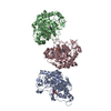
| ||||||||||||
|---|---|---|---|---|---|---|---|---|---|---|---|---|---|
| 1 | 
| ||||||||||||
| 2 | 
| ||||||||||||
| 3 | 
| ||||||||||||
| Unit cell |
|
-Components
| #1: Protein | Mass: 39511.531 Da / Num. of mol.: 3 Source method: isolated from a genetically manipulated source Source: (gene. exp.) Gluconobacter oxydans (bacteria) / Gene: nox / Production host: #2: Chemical |   Mass: 456.344 Da / Num. of mol.: 3 / Source method: obtained synthetically / Formula: C17H21N4O9P / Feature type: SUBJECT OF INVESTIGATION Mass: 456.344 Da / Num. of mol.: 3 / Source method: obtained synthetically / Formula: C17H21N4O9P / Feature type: SUBJECT OF INVESTIGATION#3: Water | ChemComp-HOH / |  Mass: 18.015 Da / Num. of mol.: 468 / Source method: isolated from a natural source / Formula: H2O Mass: 18.015 Da / Num. of mol.: 468 / Source method: isolated from a natural source / Formula: H2OHas ligand of interest | Y | |
|---|
-Experimental details
-Experiment
| Experiment | Method: X-RAY DIFFRACTION / Number of used crystals: 1 |
|---|
- Sample preparation
| Crystal | Density Matthews: 2.89 Å3/Da / Density % sol: 57.37 % |
|---|---|
| Crystal grow | Temperature: 277 K / Method: vapor diffusion, sitting drop / pH: 8 Details: 0.1 M Tris, 0.1 M potassium bromide, 20% w/v PEG4000 |
-Data collection
| Diffraction | Mean temperature: 100 K / Serial crystal experiment: N |
|---|---|
| Diffraction source | Source: SYNCHROTRON / Site: CHESS / Beamline: 7B2 / Wavelength: 0.9202 Å |
| Detector | Type: DECTRIS EIGER X 16M / Detector: PIXEL / Date: Nov 14, 2022 |
| Radiation | Protocol: SINGLE WAVELENGTH / Monochromatic (M) / Laue (L): M / Scattering type: x-ray |
| Radiation wavelength | Wavelength: 0.9202 Å / Relative weight: 1 |
| Reflection | Resolution: 1.5→45.51 Å / Num. obs: 205104 / % possible obs: 96.61 % / Redundancy: 2.5 % / Rmerge(I) obs: 0.097 / Rpim(I) all: 0.077 / Net I/σ(I): 7.9 |
| Reflection shell | Resolution: 1.5→1.53 Å / Rmerge(I) obs: 0.309 / Num. unique obs: 10107 / Rpim(I) all: 0.239 / % possible all: 93.8 |
- Processing
| Software |
| |||||||||||||||||||||||||||||||||||||||||||||||||||||||||||||||||||||||||||||||||||||||||||||||||||||||||
|---|---|---|---|---|---|---|---|---|---|---|---|---|---|---|---|---|---|---|---|---|---|---|---|---|---|---|---|---|---|---|---|---|---|---|---|---|---|---|---|---|---|---|---|---|---|---|---|---|---|---|---|---|---|---|---|---|---|---|---|---|---|---|---|---|---|---|---|---|---|---|---|---|---|---|---|---|---|---|---|---|---|---|---|---|---|---|---|---|---|---|---|---|---|---|---|---|---|---|---|---|---|---|---|---|---|---|
| Refinement | Method to determine structure: MOLECULAR REPLACEMENT Starting model: PDB entry 6MYW Resolution: 1.5→45.51 Å / Cross valid method: FREE R-VALUE / σ(F): 1.87 / Phase error: 25.5365 Stereochemistry target values: GeoStd + Monomer Library + CDL v1.2
| |||||||||||||||||||||||||||||||||||||||||||||||||||||||||||||||||||||||||||||||||||||||||||||||||||||||||
| Solvent computation | Shrinkage radii: 0.9 Å / VDW probe radii: 1.1 Å / Solvent model: FLAT BULK SOLVENT MODEL | |||||||||||||||||||||||||||||||||||||||||||||||||||||||||||||||||||||||||||||||||||||||||||||||||||||||||
| Displacement parameters | Biso mean: 16.16 Å2 | |||||||||||||||||||||||||||||||||||||||||||||||||||||||||||||||||||||||||||||||||||||||||||||||||||||||||
| Refinement step | Cycle: LAST / Resolution: 1.5→45.51 Å
| |||||||||||||||||||||||||||||||||||||||||||||||||||||||||||||||||||||||||||||||||||||||||||||||||||||||||
| Refine LS restraints |
| |||||||||||||||||||||||||||||||||||||||||||||||||||||||||||||||||||||||||||||||||||||||||||||||||||||||||
| LS refinement shell |
|
