 Movie
Movie Controller


+ Open data

- Basic information
| Entry | Database: PDB / ID: 8fux | ||||||
|---|---|---|---|---|---|---|---|
| Title | KpsC D160C ternary complex | ||||||
 Components Components | Capsule polysaccharide export protein KpsC | ||||||
 Keywords Keywords | TRANSFERASE / glycosyltransferase / retaining | ||||||
| Function / homology |  Function and homology information Function and homology information | ||||||
| Biological species |  | ||||||
| Method |  X-RAY DIFFRACTION / SYNCHROTRON / MOLECULAR REPLACEMENT / Resolution: 1.2 Å X-RAY DIFFRACTION / SYNCHROTRON / MOLECULAR REPLACEMENT / Resolution: 1.2 Å | ||||||
 Authors Authors | Kimber, M.S. / Doyle, L. / Whitfield, C. | ||||||
| Funding support |  Canada, 1items Canada, 1items
| ||||||
 Citation Citation | Journal: J.Biol.Chem. / Year: 2023 Title: Mechanism and linkage specificities of the dual retaining beta-Kdo glycosyltransferase modules of KpsC from bacterial capsule biosynthesis. Authors: Doyle, L. / Ovchinnikova, O.G. / Huang, B.S. / Forrester, T.J.B. / Lowary, T.L. / Kimber, M.S. / Whitfield, C. | ||||||
| History |
|
- Structure visualization
| Structure viewer | Molecule: MolmilJmol/JSmol |
|---|
- Downloads & links
-Download
| PDBx/mmCIF format | 8fux.cif.gz | 549.8 KB | Display | PDBx/mmCIF format |
|---|---|---|---|---|
| PDB format | pdb8fux.ent.gz | 377.4 KB | Display | PDB format |
| PDBx/mmJSON format | 8fux.json.gz | Tree view | PDBx/mmJSON format | |
| Others |  Other downloads Other downloads |
-Validation report
| Summary document | 8fux_validation.pdf.gz | 2.5 MB | Display | wwPDB validaton report |
|---|---|---|---|---|
| Full document | 8fux_full_validation.pdf.gz | 2.5 MB | Display | |
| Data in XML | 8fux_validation.xml.gz | 37.5 KB | Display | |
| Data in CIF | 8fux_validation.cif.gz | 58.7 KB | Display | |
| Arichive directory | https://data.pdbj.org/pub/pdb/validation_reports/fu/8fuxftp://data.pdbj.org/pub/pdb/validation_reports/fu/8fux | HTTPS FTP |
-Related structure data

-Links
 PDBj
PDBj- Assembly
| Deposited unit | 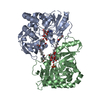
| ||||||||||||
|---|---|---|---|---|---|---|---|---|---|---|---|---|---|
| 1 | 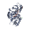
| ||||||||||||
| 2 | 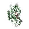
| ||||||||||||
| Unit cell |
|
-Components
-Protein / Sugars , 2 types, 4 molecules AB
| #1: Protein | Mass: 36478.633 Da / Num. of mol.: 2 / Mutation: D160C Source method: isolated from a genetically manipulated source Source: (gene. exp.) #5: Sugar |  Type: D-saccharide, alpha linking / Mass: 238.192 Da / Num. of mol.: 2 / Source method: obtained synthetically / Formula: C8H14O8 Type: D-saccharide, alpha linking / Mass: 238.192 Da / Num. of mol.: 2 / Source method: obtained synthetically / Formula: C8H14O8 |
|---|
-Non-polymers , 6 types, 947 molecules 


| #2: Chemical | ChemComp-KD3 /  Mass: 238.192 Da / Num. of mol.: 4 Mass: 238.192 Da / Num. of mol.: 4Source method: isolated from a genetically manipulated source Formula: C8H14O8 / Feature type: SUBJECT OF INVESTIGATION #3: Chemical | ChemComp-C5P / |  Mass: 323.197 Da / Num. of mol.: 1 / Source method: obtained synthetically / Formula: C9H14N3O8P / Feature type: SUBJECT OF INVESTIGATION Mass: 323.197 Da / Num. of mol.: 1 / Source method: obtained synthetically / Formula: C9H14N3O8P / Feature type: SUBJECT OF INVESTIGATION#4: Chemical |  Mass: 279.375 Da / Num. of mol.: 2 / Source method: obtained synthetically / Formula: C16H25NO3 / Feature type: SUBJECT OF INVESTIGATION Mass: 279.375 Da / Num. of mol.: 2 / Source method: obtained synthetically / Formula: C16H25NO3 / Feature type: SUBJECT OF INVESTIGATION#6: Chemical |  Mass: 35.453 Da / Num. of mol.: 2 / Source method: obtained synthetically / Formula: Cl Mass: 35.453 Da / Num. of mol.: 2 / Source method: obtained synthetically / Formula: Cl#7: Chemical | ChemComp-PO4 / |  Mass: 94.971 Da / Num. of mol.: 1 / Source method: obtained synthetically / Formula: PO4 Mass: 94.971 Da / Num. of mol.: 1 / Source method: obtained synthetically / Formula: PO4#8: Water | ChemComp-HOH / | Mass: 18.015 Da / Num. of mol.: 937 / Source method: isolated from a natural source / Formula: H2O |
|---|
-Details
| Has ligand of interest | Y |
|---|
-Experimental details
-Experiment
| Experiment | Method: X-RAY DIFFRACTION / Number of used crystals: 1 |
|---|
- Sample preparation
| Crystal | Density Matthews: 2.07 Å3/Da / Density % sol: 40.6 % |
|---|---|
| Crystal grow | Temperature: 293 K / Method: vapor diffusion, hanging drop / pH: 5.5 / Details: 25 % (v/v) PEG 3350 and 0.1 M Bis-Tris, pH 5.5 |
-Data collection
| Diffraction | Mean temperature: 100 K / Serial crystal experiment: N |
|---|---|
| Diffraction source | Source: SYNCHROTRON / Site: CLSI / Beamline: 08ID-1 / Wavelength: 0.98011 Å |
| Detector | Type: DECTRIS EIGER X 9M / Detector: PIXEL / Date: Mar 1, 2020 |
| Radiation | Protocol: SINGLE WAVELENGTH / Monochromatic (M) / Laue (L): M / Scattering type: x-ray |
| Radiation wavelength | Wavelength: 0.98011 Å / Relative weight: 1 |
| Reflection | Resolution: 1.2→48.43 Å / Num. obs: 184570 / % possible obs: 99.7 % / Redundancy: 3.3 % / Biso Wilson estimate: 12.94 Å2 / CC1/2: 0.998 / Rrim(I) all: 0.078 / Net I/σ(I): 9 |
| Reflection shell | Resolution: 1.2→1.25 Å / Redundancy: 3.2 % / Mean I/σ(I) obs: 1.39 / Num. unique obs: 21213 / CC1/2: 0.521 / Rrim(I) all: 1.104 / % possible all: 99.8 |
- Processing
| Software |
| |||||||||||||||||||||||||||||||||||||||||||||||||||||||||||||||||||||||||||||||||||||||||||||||||||||||||||||||||||||||||||||||||||||||||||||||||||||||||||||||||||||||||||||||||||||||||||||||||||||||||||||||||||||||||
|---|---|---|---|---|---|---|---|---|---|---|---|---|---|---|---|---|---|---|---|---|---|---|---|---|---|---|---|---|---|---|---|---|---|---|---|---|---|---|---|---|---|---|---|---|---|---|---|---|---|---|---|---|---|---|---|---|---|---|---|---|---|---|---|---|---|---|---|---|---|---|---|---|---|---|---|---|---|---|---|---|---|---|---|---|---|---|---|---|---|---|---|---|---|---|---|---|---|---|---|---|---|---|---|---|---|---|---|---|---|---|---|---|---|---|---|---|---|---|---|---|---|---|---|---|---|---|---|---|---|---|---|---|---|---|---|---|---|---|---|---|---|---|---|---|---|---|---|---|---|---|---|---|---|---|---|---|---|---|---|---|---|---|---|---|---|---|---|---|---|---|---|---|---|---|---|---|---|---|---|---|---|---|---|---|---|---|---|---|---|---|---|---|---|---|---|---|---|---|---|---|---|---|---|---|---|---|---|---|---|---|---|---|---|---|---|---|---|---|
| Refinement | Method to determine structure: MOLECULAR REPLACEMENT / Resolution: 1.2→48.43 Å / SU ML: 0.1339 / Cross valid method: FREE R-VALUE / σ(F): 1.34 / Phase error: 15.5116 Stereochemistry target values: GeoStd + Monomer Library + CDL v1.2
| |||||||||||||||||||||||||||||||||||||||||||||||||||||||||||||||||||||||||||||||||||||||||||||||||||||||||||||||||||||||||||||||||||||||||||||||||||||||||||||||||||||||||||||||||||||||||||||||||||||||||||||||||||||||||
| Solvent computation | Shrinkage radii: 0.9 Å / VDW probe radii: 1.11 Å / Solvent model: FLAT BULK SOLVENT MODEL | |||||||||||||||||||||||||||||||||||||||||||||||||||||||||||||||||||||||||||||||||||||||||||||||||||||||||||||||||||||||||||||||||||||||||||||||||||||||||||||||||||||||||||||||||||||||||||||||||||||||||||||||||||||||||
| Displacement parameters | Biso mean: 18.12 Å2 | |||||||||||||||||||||||||||||||||||||||||||||||||||||||||||||||||||||||||||||||||||||||||||||||||||||||||||||||||||||||||||||||||||||||||||||||||||||||||||||||||||||||||||||||||||||||||||||||||||||||||||||||||||||||||
| Refinement step | Cycle: LAST / Resolution: 1.2→48.43 Å
| |||||||||||||||||||||||||||||||||||||||||||||||||||||||||||||||||||||||||||||||||||||||||||||||||||||||||||||||||||||||||||||||||||||||||||||||||||||||||||||||||||||||||||||||||||||||||||||||||||||||||||||||||||||||||
| Refine LS restraints |
| |||||||||||||||||||||||||||||||||||||||||||||||||||||||||||||||||||||||||||||||||||||||||||||||||||||||||||||||||||||||||||||||||||||||||||||||||||||||||||||||||||||||||||||||||||||||||||||||||||||||||||||||||||||||||
| LS refinement shell |
|
