 Movie
Movie Controller

[English] 日本語
 Yorodumi
- PDB-8fuq: Crystal structure of rabies virus (strain CVS-11) P3 dimerization... -
+ Open data

- Basic information
| Entry | Database: PDB / ID: 8fuq | ||||||
|---|---|---|---|---|---|---|---|
| Title | Crystal structure of rabies virus (strain CVS-11) P3 dimerization domain | ||||||
 Components Components | Phosphoprotein | ||||||
 Keywords Keywords | VIRAL PROTEIN / rabies / phosphoprotein / lyssavirus / chaperone | ||||||
| Function / homology |  Function and homology information Function and homology information: / microtubule-dependent intracellular transport of viral material towards nucleus / viral transcription / symbiont-mediated suppression of host JAK-STAT cascade via inhibition of STAT2 activity / symbiont-mediated suppression of host JAK-STAT cascade via inhibition of STAT1 activity / virion component / host cell / symbiont-mediated suppression of host cytoplasmic pattern recognition receptor signaling pathway via inhibition of TBK1 activity / symbiont-mediated suppression of host toll-like receptor signaling pathway / host cell cytoplasm ...: / microtubule-dependent intracellular transport of viral material towards nucleus / viral transcription / symbiont-mediated suppression of host JAK-STAT cascade via inhibition of STAT2 activity / symbiont-mediated suppression of host JAK-STAT cascade via inhibition of STAT1 activity / virion component / host cell / symbiont-mediated suppression of host cytoplasmic pattern recognition receptor signaling pathway via inhibition of TBK1 activity / symbiont-mediated suppression of host toll-like receptor signaling pathway / host cell cytoplasm / symbiont-mediated suppression of host type I interferon-mediated signaling pathway / RNA-directed RNA polymerase activity / symbiont entry into host cell / host cell nucleus Similarity search - Function | ||||||
| Biological species |  Lyssavirus rabies Lyssavirus rabies | ||||||
| Method |  X-RAY DIFFRACTION / SYNCHROTRON / MOLECULAR REPLACEMENT / Resolution: 1.8 Å X-RAY DIFFRACTION / SYNCHROTRON / MOLECULAR REPLACEMENT / Resolution: 1.8 Å | ||||||
 Authors Authors | Donnelly, C.M. / Cross, E.M. / Forwood, J.K. | ||||||
| Funding support | 1items
| ||||||
 Citation Citation | Journal: To Be Published Title: Crystal structure of rabies virus (strain CVS-11) P3 dimerization domain Authors: Donnelly, C.M. / Cross, E.M. / Forwood, J.K. | ||||||
| History |
|
- Structure visualization
| Structure viewer | Molecule: MolmilJmol/JSmol |
|---|
- Downloads & links
-Download
| PDBx/mmCIF format | 8fuq.cif.gz | 197.2 KB | Display | PDBx/mmCIF format |
|---|---|---|---|---|
| PDB format | pdb8fuq.ent.gz | 152 KB | Display | PDB format |
| PDBx/mmJSON format | 8fuq.json.gz | Tree view | PDBx/mmJSON format | |
| Others |  Other downloads Other downloads |
-Validation report
| Arichive directory | https://data.pdbj.org/pub/pdb/validation_reports/fu/8fuqftp://data.pdbj.org/pub/pdb/validation_reports/fu/8fuq | HTTPS FTP |
|---|
-Related structure data
| Similar structure data |
|---|
-Links
 PDBj
PDBj
- Assembly
| Deposited unit | 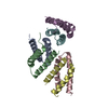
| ||||||||||||
|---|---|---|---|---|---|---|---|---|---|---|---|---|---|
| 1 | 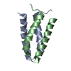
| ||||||||||||
| 2 | 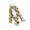
| ||||||||||||
| 3 | 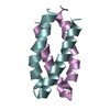
| ||||||||||||
| Unit cell |
| ||||||||||||
| Components on special symmetry positions |
|
-Components
| #1: Protein | Mass: 13737.184 Da / Num. of mol.: 6 Source method: isolated from a genetically manipulated source Source: (gene. exp.) Lyssavirus rabies / Strain: CVS-11 / Production host:  #2: Water | ChemComp-HOH / |  Mass: 18.015 Da / Num. of mol.: 169 / Source method: isolated from a natural source / Formula: H2O Mass: 18.015 Da / Num. of mol.: 169 / Source method: isolated from a natural source / Formula: H2O |
|---|
-Experimental details
-Experiment
| Experiment | Method: X-RAY DIFFRACTION / Number of used crystals: 1 |
|---|
- Sample preparation
| Crystal | Density Matthews: 0.99 Å3/Da / Density % sol: 54.62 % |
|---|---|
| Crystal grow | Temperature: 293 K / Method: vapor diffusion, hanging drop / pH: 6.5 Details: 0.2 M Sodium nitrate, 0.1 M Bis-Tris propane, 20 % w/v PEG 3350 |
-Data collection
| Diffraction | Mean temperature: 100 K / Serial crystal experiment: N |
|---|---|
| Diffraction source | Source: SYNCHROTRON / Site: Australian Synchrotron  / Beamline: MX2 / Wavelength: 0.9537 Å / Beamline: MX2 / Wavelength: 0.9537 Å |
| Detector | Type: DECTRIS EIGER2 S 16M / Detector: PIXEL / Date: Sep 15, 2020 |
| Radiation | Monochromator: M / Protocol: SINGLE WAVELENGTH / Monochromatic (M) / Laue (L): M / Scattering type: x-ray |
| Radiation wavelength | Wavelength: 0.9537 Å / Relative weight: 1 |
| Reflection | Resolution: 1.8→52.53 Å / Num. obs: 30167 / % possible obs: 99.9 % / Redundancy: 3.6 % / Biso Wilson estimate: 24.21 Å2 / CC1/2: 0.989 / Rmerge(I) obs: 0.156 / Net I/σ(I): 2.9 |
| Reflection shell | Resolution: 1.8→1.84 Å / Rmerge(I) obs: 1.521 / Mean I/σ(I) obs: 0.4 / Num. unique obs: 1797 / CC1/2: 0.248 / Rpim(I) all: 2.753 |
- Processing
| Software |
| ||||||||||||||||||||||||||||||||||||||||||||||||||||||||||||||||||||||||||||||||||||
|---|---|---|---|---|---|---|---|---|---|---|---|---|---|---|---|---|---|---|---|---|---|---|---|---|---|---|---|---|---|---|---|---|---|---|---|---|---|---|---|---|---|---|---|---|---|---|---|---|---|---|---|---|---|---|---|---|---|---|---|---|---|---|---|---|---|---|---|---|---|---|---|---|---|---|---|---|---|---|---|---|---|---|---|---|---|
| Refinement | Method to determine structure: MOLECULAR REPLACEMENT / Resolution: 1.8→52.51 Å / SU ML: 0.2774 / Cross valid method: FREE R-VALUE / σ(F): 1.33 / Phase error: 26.5342 Stereochemistry target values: GeoStd + Monomer Library + CDL v1.2
| ||||||||||||||||||||||||||||||||||||||||||||||||||||||||||||||||||||||||||||||||||||
| Solvent computation | Shrinkage radii: 0.9 Å / VDW probe radii: 1.1 Å / Solvent model: FLAT BULK SOLVENT MODEL | ||||||||||||||||||||||||||||||||||||||||||||||||||||||||||||||||||||||||||||||||||||
| Displacement parameters | Biso mean: 31.76 Å2 | ||||||||||||||||||||||||||||||||||||||||||||||||||||||||||||||||||||||||||||||||||||
| Refinement step | Cycle: LAST / Resolution: 1.8→52.51 Å
| ||||||||||||||||||||||||||||||||||||||||||||||||||||||||||||||||||||||||||||||||||||
| Refine LS restraints |
| ||||||||||||||||||||||||||||||||||||||||||||||||||||||||||||||||||||||||||||||||||||
| LS refinement shell |
|
