 Movie
Movie Controller


+ Open data

- Basic information
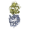
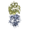
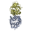
| Entry | Database: PDB / ID: 8fu5 | ||||||
|---|---|---|---|---|---|---|---|
| Title | Crystal structure of Fe-CAO1 in complex with piceatannol | ||||||
 Components Components | Carotenoid oxygenase | ||||||
 Keywords Keywords | OXIDOREDUCTASE/SUBSTRATE / non-heme iron / beta propeller / stilbene / dioxygenase / oxidoreductase / OXIDOREDUCTASE-SUBSTRATE complex | ||||||
| Function / homology | Carotenoid oxygenase / Retinal pigment epithelial membrane protein / oxidoreductase activity, acting on single donors with incorporation of molecular oxygen, incorporation of two atoms of oxygen / metal ion binding / BENZOIC ACID / : / PICEATANNOL / Carotenoid oxygenase Function and homology information Function and homology information | ||||||
| Biological species |  Neurospora crassa (fungus) Neurospora crassa (fungus) | ||||||
| Method |  X-RAY DIFFRACTION / SYNCHROTRON / FOURIER SYNTHESIS / Resolution: 2.01 Å X-RAY DIFFRACTION / SYNCHROTRON / FOURIER SYNTHESIS / Resolution: 2.01 Å | ||||||
 Authors Authors | Kiser, P.D. | ||||||
| Funding support |  United States, 1items United States, 1items
| ||||||
 Citation Citation | Journal: J.Biol.Chem. / Year: 2025 Title: Spectroscopy and crystallography define carotenoid oxygenases as a new subclass of mononuclear non-heme Fe II enzymes. Authors: DeWeese, D.E. / Everett, M.P. / Babicz Jr., J.T. / Daruwalla, A. / Solomon, E.I. / Kiser, P.D. | ||||||
| History |
|
- Structure visualization
| Structure viewer | Molecule: MolmilJmol/JSmol |
|---|
- Downloads & links
-Download
| PDBx/mmCIF format | 8fu5.cif.gz | 452.4 KB | Display | PDBx/mmCIF format |
|---|---|---|---|---|
| PDB format | pdb8fu5.ent.gz | 365.2 KB | Display | PDB format |
| PDBx/mmJSON format | 8fu5.json.gz | Tree view | PDBx/mmJSON format | |
| Others |  Other downloads Other downloads |
-Validation report
| Arichive directory | https://data.pdbj.org/pub/pdb/validation_reports/fu/8fu5ftp://data.pdbj.org/pub/pdb/validation_reports/fu/8fu5 | HTTPS FTP |
|---|
-Related structure data
-Links
 PDBj
PDBj


- Assembly
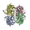
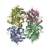
| Deposited unit | 
| ||||||||
|---|---|---|---|---|---|---|---|---|---|
| 1 | 
| ||||||||
| 2 | 
| ||||||||
| Unit cell |
|
-Components
| #1: Protein | Mass: 59497.934 Da / Num. of mol.: 4 Source method: isolated from a genetically manipulated source Source: (gene. exp.) Neurospora crassa (fungus) / Gene: GE21DRAFT_6499 / Production host:  #2: Chemical | ChemComp-FE2 /   Mass: 55.845 Da / Num. of mol.: 4 / Source method: obtained synthetically / Formula: Fe / Feature type: SUBJECT OF INVESTIGATION Mass: 55.845 Da / Num. of mol.: 4 / Source method: obtained synthetically / Formula: Fe / Feature type: SUBJECT OF INVESTIGATION#3: Chemical | ChemComp-PIT / 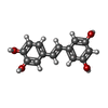  Mass: 244.243 Da / Num. of mol.: 4 / Source method: obtained synthetically / Formula: C14H12O4 / Feature type: SUBJECT OF INVESTIGATION Mass: 244.243 Da / Num. of mol.: 4 / Source method: obtained synthetically / Formula: C14H12O4 / Feature type: SUBJECT OF INVESTIGATION#4: Chemical | ChemComp-BEZ /   Mass: 122.121 Da / Num. of mol.: 4 / Source method: obtained synthetically / Formula: C7H6O2 Mass: 122.121 Da / Num. of mol.: 4 / Source method: obtained synthetically / Formula: C7H6O2#5: Water | ChemComp-HOH / |  Mass: 18.015 Da / Num. of mol.: 2023 / Source method: isolated from a natural source / Formula: H2O Mass: 18.015 Da / Num. of mol.: 2023 / Source method: isolated from a natural source / Formula: H2OHas ligand of interest | Y | Has protein modification | N | |
|---|
-Experimental details
-Experiment
| Experiment | Method: X-RAY DIFFRACTION / Number of used crystals: 1 |
|---|
- Sample preparation
| Crystal | Density Matthews: 2.79 Å3/Da / Density % sol: 55.92 % |
|---|---|
| Crystal grow | Temperature: 277.15 K / Method: vapor diffusion, hanging drop / pH: 7 Details: 0.1 M HEPES-NaOH pH 7, 42% sodium polyacrylate 2100 |
-Data collection
| Diffraction | Mean temperature: 100 K / Serial crystal experiment: N |
|---|---|
| Diffraction source | Source: SYNCHROTRON / Site: APS / Beamline: 24-ID-E / Wavelength: 0.97918 Å |
| Detector | Type: DECTRIS EIGER X 16M / Detector: PIXEL / Date: Sep 29, 2022 |
| Radiation | Protocol: SINGLE WAVELENGTH / Monochromatic (M) / Laue (L): M / Scattering type: x-ray |
| Radiation wavelength | Wavelength: 0.97918 Å / Relative weight: 1 |
| Reflection | Resolution: 2.01→50 Å / Num. obs: 179310 / % possible obs: 99.8 % / Redundancy: 6.8 % / CC1/2: 0.996 / Rmerge(I) obs: 0.234 / Net I/σ(I): 10.54 |
| Reflection shell | Resolution: 2.01→2.13 Å / Redundancy: 6.8 % / Rmerge(I) obs: 2.067 / Mean I/σ(I) obs: 1.18 / Num. unique obs: 28387 / CC1/2: 0.456 / % possible all: 99.1 |
- Processing
| Software |
| ||||||||||||||||||||||||||||||||||||||||||||||||||||||||||||||||||||||||||||||||||||||||||||||||||||||||||||||||||||||||||||||||||||||||||||||||||||||||||||||||||||||||||||||||||||||
|---|---|---|---|---|---|---|---|---|---|---|---|---|---|---|---|---|---|---|---|---|---|---|---|---|---|---|---|---|---|---|---|---|---|---|---|---|---|---|---|---|---|---|---|---|---|---|---|---|---|---|---|---|---|---|---|---|---|---|---|---|---|---|---|---|---|---|---|---|---|---|---|---|---|---|---|---|---|---|---|---|---|---|---|---|---|---|---|---|---|---|---|---|---|---|---|---|---|---|---|---|---|---|---|---|---|---|---|---|---|---|---|---|---|---|---|---|---|---|---|---|---|---|---|---|---|---|---|---|---|---|---|---|---|---|---|---|---|---|---|---|---|---|---|---|---|---|---|---|---|---|---|---|---|---|---|---|---|---|---|---|---|---|---|---|---|---|---|---|---|---|---|---|---|---|---|---|---|---|---|---|---|---|---|
| Refinement | Method to determine structure: FOURIER SYNTHESIS / Resolution: 2.01→49.36 Å / Cor.coef. Fo:Fc: 0.969 / Cor.coef. Fo:Fc free: 0.958 / SU B: 4.756 / SU ML: 0.118 / Cross valid method: THROUGHOUT / ESU R: 0.15 / ESU R Free: 0.131 / Stereochemistry target values: MAXIMUM LIKELIHOOD / Details: HYDROGENS HAVE BEEN ADDED IN THE RIDING POSITIONS
| ||||||||||||||||||||||||||||||||||||||||||||||||||||||||||||||||||||||||||||||||||||||||||||||||||||||||||||||||||||||||||||||||||||||||||||||||||||||||||||||||||||||||||||||||||||||
| Solvent computation | Ion probe radii: 0.7 Å / Shrinkage radii: 0.7 Å / VDW probe radii: 1.1 Å / Solvent model: MASK | ||||||||||||||||||||||||||||||||||||||||||||||||||||||||||||||||||||||||||||||||||||||||||||||||||||||||||||||||||||||||||||||||||||||||||||||||||||||||||||||||||||||||||||||||||||||
| Displacement parameters | Biso mean: 30.755 Å2
| ||||||||||||||||||||||||||||||||||||||||||||||||||||||||||||||||||||||||||||||||||||||||||||||||||||||||||||||||||||||||||||||||||||||||||||||||||||||||||||||||||||||||||||||||||||||
| Refinement step | Cycle: 1 / Resolution: 2.01→49.36 Å
| ||||||||||||||||||||||||||||||||||||||||||||||||||||||||||||||||||||||||||||||||||||||||||||||||||||||||||||||||||||||||||||||||||||||||||||||||||||||||||||||||||||||||||||||||||||||
| Refine LS restraints |
|