- PDB-8ftj: Crystal structure of human NEIL1 (P2G (242K) C(delta)100) glycosy... -
+
Open data
ID or keywords:
Loading...
-
Basic information
Entry
Database: PDB / ID: 8ftj
Title
Crystal structure of human NEIL1 (P2G (242K) C(delta)100) glycosylase bound to DNA duplex containing urea
Components
DNA (5'-D(*CP*GP*TP*CP*CP*AP*UDV*GP*TP*CP*TP*AP*CP)-3')
DNA (5'-D(*TP*AP*GP*AP*CP*AP*TP*GP*GP*AP*CP*GP*G)-3')
Endonuclease 8-like 1
Keywords
DNA BINDING PROTEIN/DNA / base-excision repair enzyme / DNA glycosylase / DNA binding protein / DNA binding protein-DNA complex
Function / homology
Function and homology information
: / Defective Base Excision Repair Associated with NEIL1 / depyrimidination / DNA N-glycosylase activity / hydrolase activity, acting on glycosyl bonds / Hydrolases; Glycosylases; Hydrolysing N-glycosyl compounds / APEX1-Independent Resolution of AP Sites via the Single Nucleotide Replacement Pathway / base-excision repair, gap-filling / DNA-(apurinic or apyrimidinic site) endonuclease activity / Recognition and association of DNA glycosylase with site containing an affected pyrimidine ...: / Defective Base Excision Repair Associated with NEIL1 / depyrimidination / DNA N-glycosylase activity / hydrolase activity, acting on glycosyl bonds / Hydrolases; Glycosylases; Hydrolysing N-glycosyl compounds / APEX1-Independent Resolution of AP Sites via the Single Nucleotide Replacement Pathway / base-excision repair, gap-filling / DNA-(apurinic or apyrimidinic site) endonuclease activity / Recognition and association of DNA glycosylase with site containing an affected pyrimidine / Cleavage of the damaged pyrimidine / DNA-(apurinic or apyrimidinic site) lyase / class I DNA-(apurinic or apyrimidinic site) endonuclease activity / base-excision repair / chromosome / response to oxidative stress / damaged DNA binding / centrosome / zinc ion binding / nucleoplasm / nucleus / cytosol / cytoplasm Similarity search - Function
Endonuclease8-like1 / DNA glycosylase/AP lyase Neil1 / DNA-(apurinic or apyrimidinic site) lyase Neil1 / Endonuclease ...DNA glycosylase/AP lyase Neil1 / DNA-(apurinic or apyrimidinic site) lyase Neil1 / Endonuclease VIII-like 1 / FPG1 / Nei homolog 1 / NEH1 / Nei-like protein 1
Mass: 33641.414 Da / Num. of mol.: 1 / Mutation: P2G Source method: isolated from a genetically manipulated source Source: (gene. exp.) Homo sapiens (human) / Gene: NEIL1 / Production host: Escherichia coli (E. coli) References: UniProt: Q96FI4, Hydrolases; Glycosylases; Hydrolysing N-glycosyl compounds, DNA-(apurinic or apyrimidinic site) lyase
-
DNA chain , 2 types, 2 molecules DF
#2: DNA chain
DNA (5'-D(*CP*GP*TP*CP*CP*AP*UDV*GP*TP*CP*TP*AP*CP)-3')
Mass: 3838.506 Da / Num. of mol.: 1 / Source method: obtained synthetically Details: YB9 is modeled into the chain D of DNA template strand at position 297; basically urea moiety is covalently linked to ring-opened deoxyribose in YB9 residue ie. YB9 is modified nucleotide in ...Details: YB9 is modeled into the chain D of DNA template strand at position 297; basically urea moiety is covalently linked to ring-opened deoxyribose in YB9 residue ie. YB9 is modified nucleotide in DNA template strand (Chain D) Source: (synth.) synthetic construct (others)
#3: DNA chain
DNA (5'-D(*TP*AP*GP*AP*CP*AP*TP*GP*GP*AP*CP*GP*G)-3')
Mass: 4040.647 Da / Num. of mol.: 1 / Source method: obtained synthetically Details: Complementary DNA strand (Chain F) to template strand (chain D) Source: (synth.) synthetic construct (others)
Mass: 18.015 Da / Num. of mol.: 127 / Source method: isolated from a natural source / Formula: H2O
-
Details
Has ligand of interest
Y
Has protein modification
Y
-
Experimental details
-
Experiment
Experiment
Method: X-RAY DIFFRACTION / Number of used crystals: 1
-
Sample preparation
Crystal
Density Matthews: 2.73 Å3/Da / Density % sol: 54.9 %
Crystal grow
Temperature: 287 K / Method: vapor diffusion, hanging drop Details: 0.01 M magnesium sulfate and 1.8 M lithium sulfate, 0.05 M sodium cacodylate, pH 6.0
-
Data collection
Diffraction
Mean temperature: 100 K / Serial crystal experiment: N
In the structure databanks used in Yorodumi, some data are registered as the other names, "COVID-19 virus" and "2019-nCoV". Here are the details of the virus and the list of structure data.
Jan 31, 2019. EMDB accession codes are about to change! (news from PDBe EMDB page)
EMDB accession codes are about to change! (news from PDBe EMDB page)
The allocation of 4 digits for EMDB accession codes will soon come to an end. Whilst these codes will remain in use, new EMDB accession codes will include an additional digit and will expand incrementally as the available range of codes is exhausted. The current 4-digit format prefixed with “EMD-” (i.e. EMD-XXXX) will advance to a 5-digit format (i.e. EMD-XXXXX), and so on. It is currently estimated that the 4-digit codes will be depleted around Spring 2019, at which point the 5-digit format will come into force.
The EM Navigator/Yorodumi systems omit the EMD- prefix.
Related info.:Q: What is EMD? / ID/Accession-code notation in Yorodumi/EM Navigator
Yorodumi is a browser for structure data from EMDB, PDB, SASBDB, etc.
This page is also the successor to EM Navigator detail page, and also detail information page/front-end page for Omokage search.
The word "yorodu" (or yorozu) is an old Japanese word meaning "ten thousand". "mi" (miru) is to see.
Related info.:EMDB / PDB / SASBDB / Comparison of 3 databanks / Yorodumi Search / Aug 31, 2016. New EM Navigator & Yorodumi / Yorodumi Papers / Jmol/JSmol / Function and homology information / Changes in new EM Navigator and Yorodumi
 Movie
Movie Controller

 Yorodumi
 Open data
 Basic information
 Components
 Keywords
 Function and homology information
 Homo sapiens (human)
 X-RAY DIFFRACTION /
  Authors
 United States, 1items
  Citation
 Structure visualization
 Downloads & links
 Other downloads
 PDBj
PDBj


 Assembly
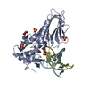


 Mass: 62.068 Da / Num. of mol.: 3 / Source method: obtained synthetically / Formula: C2H6O2 / Feature type: SUBJECT OF INVESTIGATION
 Mass: 96.063 Da / Num. of mol.: 3 / Source method: obtained synthetically / Formula: SO4 / Feature type: SUBJECT OF INVESTIGATION
 Sample preparation
 Processing