 Movie
Movie Controller


+ Open data

- Basic information
| Entry | Database: PDB / ID: 8ftf | ||||||
|---|---|---|---|---|---|---|---|
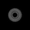
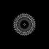
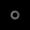
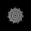
| Title | CryoEM strucutre of 33-mer RBM3 of the Salmonella MS-ring | ||||||
 Components Components | Flagellar M-ring protein | ||||||
 Keywords Keywords | MOTOR PROTEIN / flagellar / 33-fold / ring-building motif / MS-ring / FliF | ||||||
| Function / homology |  Function and homology information Function and homology informationbacterial-type flagellum basal body, MS ring / cytoskeletal motor activity / bacterial-type flagellum-dependent cell motility / plasma membrane Similarity search - Function | ||||||
| Biological species |  Salmonella enterica subsp. enterica serovar Typhimurium (bacteria) Salmonella enterica subsp. enterica serovar Typhimurium (bacteria) | ||||||
| Method | ELECTRON MICROSCOPY / single particle reconstruction / cryo EM / Resolution: 2.9 Å | ||||||
 Authors Authors | Singh, P.S. / Nakagawa, T. / Cecchini, G. / Iverson, T.M. | ||||||
| Funding support |  United States, 1items United States, 1items
| ||||||
 Citation Citation | Journal: PLoS One / Year: 2023 Title: CryoEM structure of a post-assembly MS-ring reveals plasticity in stoichiometry and conformation. Authors: Prashant K Singh / Gary Cecchini / Terunaga Nakagawa / T M Iverson / Abstract: The flagellar motor supports bacterial chemotaxis, a process that allows bacteria to move in response to their environment. A central feature of this motor is the MS-ring, which is composed entirely ...The flagellar motor supports bacterial chemotaxis, a process that allows bacteria to move in response to their environment. A central feature of this motor is the MS-ring, which is composed entirely of repeats of the FliF subunit. This MS-ring is critical for the assembly and stability of the flagellar switch and the entire flagellum. Despite multiple independent cryoEM structures of the MS-ring, there remains a debate about the stoichiometry and organization of the ring-building motifs (RBMs). Here, we report the cryoEM structure of a Salmonella MS-ring that was purified from the assembled flagellar switch complex (MSC-ring). We term this the 'post-assembly' state. Using 2D class averages, we show that under these conditions, the post-assembly MS-ring can contain 32, 33, or 34 FliF subunits, with 33 being the most common. RBM3 has a single location with C32, C33, or C34 symmetry. RBM2 is found in two locations with RBM2inner having C21 or C22 symmetry and an RBM2outer-RBM1 having C11 symmetry. Comparison to previously reported structures identifies several differences. Most strikingly, we find that the membrane domain forms 11 regions of discrete density at the base of the structure rather than a contiguous ring, although density could not be unambiguously interpreted. We further find density in some previously unresolved areas, and we assigned amino acids to those regions. Finally, we find differences in interdomain angles in RBM3 that affect the diameter of the ring. Together, these investigations support a model of the flagellum with structural plasticity, which may be important for flagellar assembly and function. | ||||||
| History |
|
- Structure visualization
| Structure viewer | Molecule: MolmilJmol/JSmol |
|---|
- Downloads & links
-Download
| PDBx/mmCIF format | 8ftf.cif.gz | 1 MB | Display | PDBx/mmCIF format |
|---|---|---|---|---|
| PDB format | pdb8ftf.ent.gz | Display | PDB format | |
| PDBx/mmJSON format | 8ftf.json.gz | Tree view | PDBx/mmJSON format | |
| Others |  Other downloads Other downloads |
-Validation report
| Arichive directory | https://data.pdbj.org/pub/pdb/validation_reports/ft/8ftfftp://data.pdbj.org/pub/pdb/validation_reports/ft/8ftf | HTTPS FTP |
|---|
-Related structure data
| Related structure data | 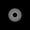 29425MC 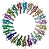 8fteC M: map data used to model this data C: citing same article ( |
|---|---|
| Similar structure data |
-Links
 PDBj
PDBj
- Assembly
| Deposited unit | 
|
|---|---|
| 1 |
|
-Components
| #1: Protein | Mass: 61295.645 Da / Num. of mol.: 33 Source method: isolated from a genetically manipulated source Source: (gene. exp.) Salmonella enterica subsp. enterica serovar Typhimurium (bacteria)Gene: fliF, fla AII.1, fla BI, STM1969 / Production host: |
|---|
-Experimental details
-Experiment
| Experiment | Method: ELECTRON MICROSCOPY |
|---|---|
| EM experiment | Aggregation state: PARTICLE / 3D reconstruction method: single particle reconstruction |
- Sample preparation
| Component | Name: MS-ring of the flagellar complex / Type: COMPLEX Details: CryoEM strucutre of the 33-mer RBM3 region of the Salmonella MS-ring Entity ID: all / Source: RECOMBINANT | ||||||||||||||||||||
|---|---|---|---|---|---|---|---|---|---|---|---|---|---|---|---|---|---|---|---|---|---|
| Molecular weight | Value: 2.5 MDa / Experimental value: NO | ||||||||||||||||||||
| Source (natural) | Organism: Salmonella enterica subsp. enterica serovar Typhimurium (bacteria) | ||||||||||||||||||||
| Source (recombinant) | Organism: | ||||||||||||||||||||
| Buffer solution | pH: 7.5 Details: 50 mM Tris-HCl pH 7.5, 100 mM NaCl, 0.1 % Ana-grade LDAO | ||||||||||||||||||||
| Buffer component |
| ||||||||||||||||||||
| Specimen | Embedding applied: NO / Shadowing applied: NO / Staining applied: NO / Vitrification applied: YES | ||||||||||||||||||||
| Specimen support | Details: Quantifoil grid coated with graphene oxide substrate (Electron Microscopy Sciences) was glow discharged for 5 sec. Purified MS-rings were added to each grid at 4C at 100% humidity. After 30 ...Details: Quantifoil grid coated with graphene oxide substrate (Electron Microscopy Sciences) was glow discharged for 5 sec. Purified MS-rings were added to each grid at 4C at 100% humidity. After 30 sec of incubation, blotting was performed for 12 sec. The grid was then plunged into liquid ethane using a Vitrobot Mark IV system (Thermo Fisher). Grid material: GRAPHENE OXIDE / Grid mesh size: 400 divisions/in. / Grid type: UltrAuFoil R1.2/1.3 | ||||||||||||||||||||
| Vitrification | Instrument: FEI VITROBOT MARK IV / Cryogen name: ETHANE / Humidity: 100 % / Chamber temperature: 277.15 K Details: Purified MS-rings were added to each grid at 277.15K at 100% humidity. After 30 sec of incubation, blotting was performed for 12 sec. The grid was then plunged into liquid ethane using a ...Details: Purified MS-rings were added to each grid at 277.15K at 100% humidity. After 30 sec of incubation, blotting was performed for 12 sec. The grid was then plunged into liquid ethane using a Vitrobot Mark IV system (Thermo Fisher) |
- Electron microscopy imaging
| Experimental equipment |  Model: Titan Krios / Image courtesy: FEI Company |
|---|---|
| Microscopy | Model: FEI TITAN KRIOS |
| Electron gun | Electron source:  FIELD EMISSION GUN / Accelerating voltage: 300 kV / Illumination mode: FLOOD BEAM FIELD EMISSION GUN / Accelerating voltage: 300 kV / Illumination mode: FLOOD BEAM |
| Electron lens | Mode: BRIGHT FIELD / Nominal defocus max: 2200 nm / Nominal defocus min: 800 nm |
| Image recording | Electron dose: 50 e/Å2 / Film or detector model: FEI FALCON III (4k x 4k) |
- Processing
| EM software |
| ||||||||||||||||||||||||||||||||||||
|---|---|---|---|---|---|---|---|---|---|---|---|---|---|---|---|---|---|---|---|---|---|---|---|---|---|---|---|---|---|---|---|---|---|---|---|---|---|
| CTF correction | Type: PHASE FLIPPING AND AMPLITUDE CORRECTION | ||||||||||||||||||||||||||||||||||||
| Symmetry | Point symmetry: C33 (33 fold cyclic) | ||||||||||||||||||||||||||||||||||||
| 3D reconstruction | Resolution: 2.9 Å / Resolution method: FSC 0.143 CUT-OFF / Num. of particles: 32042 / Symmetry type: POINT |
