 Movie
Movie Controller


+ Open data

- Basic information
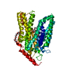
| Entry | Database: PDB / ID: 8frh | ||||||
|---|---|---|---|---|---|---|---|
| Title | Apo structure of D59C mutant of a melibiose transporter | ||||||
 Components Components | Melibiose permease | ||||||
 Keywords Keywords | MEMBRANE PROTEIN / sugar transporter / Na-coupled MFS symporter | ||||||
| Function / homology |  Function and homology information Function and homology informationsymporter activity / sodium ion transport / carbohydrate transport / transmembrane transport / plasma membrane Similarity search - Function | ||||||
| Biological species |  Salmonella enterica subsp. enterica serovar Typhimurium str. LT2 (bacteria) Salmonella enterica subsp. enterica serovar Typhimurium str. LT2 (bacteria) | ||||||
| Method |  X-RAY DIFFRACTION / SYNCHROTRON / MOLECULAR REPLACEMENT / Resolution: 3.18 Å X-RAY DIFFRACTION / SYNCHROTRON / MOLECULAR REPLACEMENT / Resolution: 3.18 Å | ||||||
 Authors Authors | Guan, L. / Hariharan, P. | ||||||
| Funding support |  United States, 1items United States, 1items
| ||||||
 Citation Citation | Journal: J.Biol.Chem. / Year: 2024 Title: Distinct roles of the major binding residues in the cation-binding pocket of the melibiose transporter MelB. Authors: Hariharan, P. / Bakhtiiari, A. / Liang, R. / Guan, L. | ||||||
| History |
|
- Structure visualization
| Structure viewer | Molecule: MolmilJmol/JSmol |
|---|
- Downloads & links
-Download
| PDBx/mmCIF format | 8frh.cif.gz | 325.8 KB | Display | PDBx/mmCIF format |
|---|---|---|---|---|
| PDB format | pdb8frh.ent.gz | 226.4 KB | Display | PDB format |
| PDBx/mmJSON format | 8frh.json.gz | Tree view | PDBx/mmJSON format | |
| Others |  Other downloads Other downloads |
-Validation report
| Arichive directory | https://data.pdbj.org/pub/pdb/validation_reports/fr/8frhftp://data.pdbj.org/pub/pdb/validation_reports/fr/8frh | HTTPS FTP |
|---|
-Related structure data
-Links
 PDBj
PDBj


- Assembly
| Deposited unit | 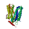
| ||||||||||||
|---|---|---|---|---|---|---|---|---|---|---|---|---|---|
| 1 |
| ||||||||||||
| Unit cell |
|
-Components
| #1: Protein | Mass: 54092.496 Da / Num. of mol.: 1 Source method: isolated from a genetically manipulated source Source: (gene. exp.) Salmonella enterica subsp. enterica serovar Typhimurium str. LT2 (bacteria)Strain: LT2 / Gene: melB, STM4299 / Production host: |
|---|---|
| #2: Chemical | ChemComp-1PE /   Mass: 238.278 Da / Num. of mol.: 1 / Source method: obtained synthetically / Formula: C10H22O6 / Feature type: SUBJECT OF INVESTIGATION / Comment: precipitant*YM Mass: 238.278 Da / Num. of mol.: 1 / Source method: obtained synthetically / Formula: C10H22O6 / Feature type: SUBJECT OF INVESTIGATION / Comment: precipitant*YM |
| Has ligand of interest | Y |
| Has protein modification | N |
-Experimental details
-Experiment
| Experiment | Method: X-RAY DIFFRACTION / Number of used crystals: 1 |
|---|
- Sample preparation
| Crystal | Density Matthews: 4.56 Å3/Da / Density % sol: 73.05 % |
|---|---|
| Crystal grow | Temperature: 296.15 K / Method: vapor diffusion, hanging drop / pH: 8.5 Details: 100 mM Tris-HCl, pH 8.5 100 mm NaCl 50 mM CaCL2 32% PEG400 |
-Data collection
| Diffraction | Mean temperature: 100 K / Serial crystal experiment: N |
|---|---|
| Diffraction source | Source: SYNCHROTRON / Site: ALS / Beamline: 8.2.2 / Wavelength: 1 Å |
| Detector | Type: ADSC QUANTUM 315r / Detector: CCD / Date: Jul 1, 2021 |
| Radiation | Protocol: SINGLE WAVELENGTH / Monochromatic (M) / Laue (L): M / Scattering type: x-ray |
| Radiation wavelength | Wavelength: 1 Å / Relative weight: 1 |
| Reflection | Resolution: 3.18→20 Å / Num. obs: 16869 / % possible obs: 99.9 % / Observed criterion σ(I): 2 / Redundancy: 21.8 % / CC1/2: 1 / Rmerge(I) obs: 0.149 / Rpim(I) all: 0.037 / Χ2: 1.3 / Net I/σ(I): 21.2 |
| Reflection shell | Resolution: 3.18→3.27 Å / Redundancy: 22.3 % / Rmerge(I) obs: 0.149 / Mean I/σ(I) obs: 21.2 / Num. unique obs: 16869 / CC1/2: 0.436 / Rpim(I) all: 0.451 / Rrim(I) all: 0.156 / Χ2: 1.3 / % possible all: 99.5 |
- Processing
| Software |
| |||||||||||||||||||||||||||||||||||||||||||||||||||||||||||||||||||||||||||||||||||||||||||
|---|---|---|---|---|---|---|---|---|---|---|---|---|---|---|---|---|---|---|---|---|---|---|---|---|---|---|---|---|---|---|---|---|---|---|---|---|---|---|---|---|---|---|---|---|---|---|---|---|---|---|---|---|---|---|---|---|---|---|---|---|---|---|---|---|---|---|---|---|---|---|---|---|---|---|---|---|---|---|---|---|---|---|---|---|---|---|---|---|---|---|---|---|
| Refinement | Method to determine structure: MOLECULAR REPLACEMENT / Resolution: 3.18→19.91 Å / SU ML: 0.5017 / Cross valid method: FREE R-VALUE / σ(F): 1.34 / Phase error: 32.7163 Stereochemistry target values: GeoStd + Monomer Library + CDL v1.2
| |||||||||||||||||||||||||||||||||||||||||||||||||||||||||||||||||||||||||||||||||||||||||||
| Solvent computation | Shrinkage radii: 0.9 Å / VDW probe radii: 1.11 Å / Solvent model: FLAT BULK SOLVENT MODEL | |||||||||||||||||||||||||||||||||||||||||||||||||||||||||||||||||||||||||||||||||||||||||||
| Displacement parameters | Biso mean: 115.05 Å2 | |||||||||||||||||||||||||||||||||||||||||||||||||||||||||||||||||||||||||||||||||||||||||||
| Refinement step | Cycle: LAST / Resolution: 3.18→19.91 Å
| |||||||||||||||||||||||||||||||||||||||||||||||||||||||||||||||||||||||||||||||||||||||||||
| Refine LS restraints |
| |||||||||||||||||||||||||||||||||||||||||||||||||||||||||||||||||||||||||||||||||||||||||||
| LS refinement shell |
| |||||||||||||||||||||||||||||||||||||||||||||||||||||||||||||||||||||||||||||||||||||||||||
| Refinement TLS params. | Method: refined / Origin x: -25.2442813874 Å / Origin y: 50.8654464875 Å / Origin z: -6.22304196232 Å
| |||||||||||||||||||||||||||||||||||||||||||||||||||||||||||||||||||||||||||||||||||||||||||
| Refinement TLS group | Selection details: all |
