 Movie
Movie Controller


+ Open data

- Basic information
| Entry | Database: PDB / ID: 8fmm | ||||||
|---|---|---|---|---|---|---|---|
| Title | Complex structure of wild type Troponin complex | ||||||
 Components Components |
| ||||||
 Keywords Keywords | STRUCTURAL PROTEIN / Ca sensing complex / troponin complex | ||||||
| Function / homology |  Function and homology information Function and homology informationregulation of systemic arterial blood pressure by ischemic conditions / troponin C binding / diaphragm contraction / regulation of ATP-dependent activity / regulation of muscle filament sliding speed / troponin T binding / cardiac Troponin complex / cardiac myofibril / troponin complex / regulation of muscle contraction ...regulation of systemic arterial blood pressure by ischemic conditions / troponin C binding / diaphragm contraction / regulation of ATP-dependent activity / regulation of muscle filament sliding speed / troponin T binding / cardiac Troponin complex / cardiac myofibril / troponin complex / regulation of muscle contraction / regulation of smooth muscle contraction / negative regulation of ATP-dependent activity / transition between fast and slow fiber / positive regulation of ATP-dependent activity / Striated Muscle Contraction / muscle filament sliding / response to metal ion / regulation of cardiac muscle contraction by calcium ion signaling / ventricular cardiac muscle tissue morphogenesis / heart contraction / regulation of heart contraction / tropomyosin binding / troponin I binding / striated muscle thin filament / skeletal muscle contraction / calcium channel inhibitor activity / vasculogenesis / Ion homeostasis / cardiac muscle contraction / sarcomere / intracellular calcium ion homeostasis / response to calcium ion / calcium-dependent protein binding / actin filament binding / actin binding / heart development / protein domain specific binding / calcium ion binding / protein kinase binding / protein homodimerization activity / identical protein binding / cytosol Similarity search - Function | ||||||
| Biological species |  Homo sapiens (human) Homo sapiens (human) | ||||||
| Method |  X-RAY DIFFRACTION / SYNCHROTRON / MOLECULAR REPLACEMENT / Resolution: 3.112 Å X-RAY DIFFRACTION / SYNCHROTRON / MOLECULAR REPLACEMENT / Resolution: 3.112 Å | ||||||
 Authors Authors | Wang, P. / Ahmed, M. / Sadek, H. | ||||||
| Funding support |  United States, 1items United States, 1items
| ||||||
 Citation Citation | Journal: To Be Published Title: Structural and Phenotypic Correction of K210del Genetic Cardiomyopathy by an FDA Approved drug Authors: Wang, P. / Ahmed, M. / Sadek, H. | ||||||
| History |
|
- Structure visualization
| Structure viewer | Molecule: MolmilJmol/JSmol |
|---|
- Downloads & links
-Download
| PDBx/mmCIF format | 8fmm.cif.gz | 148.4 KB | Display | PDBx/mmCIF format |
|---|---|---|---|---|
| PDB format | pdb8fmm.ent.gz | 114.7 KB | Display | PDB format |
| PDBx/mmJSON format | 8fmm.json.gz | Tree view | PDBx/mmJSON format | |
| Others |  Other downloads Other downloads |
-Validation report
| Summary document | 8fmm_validation.pdf.gz | 2.1 MB | Display | wwPDB validaton report |
|---|---|---|---|---|
| Full document | 8fmm_full_validation.pdf.gz | 2.1 MB | Display | |
| Data in XML | 8fmm_validation.xml.gz | 24 KB | Display | |
| Data in CIF | 8fmm_validation.cif.gz | 32.5 KB | Display | |
| Arichive directory | https://data.pdbj.org/pub/pdb/validation_reports/fm/8fmmftp://data.pdbj.org/pub/pdb/validation_reports/fm/8fmm | HTTPS FTP |
-Related structure data
| Related structure data | 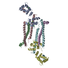 8fmnC  8fmoC  8fmpC  8fmqC 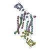 8fmrC 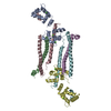 8fmsC  8fmtC C: citing same article ( |
|---|---|
| Similar structure data |
-Links
 PDBj
PDBj


- Assembly
| Deposited unit | 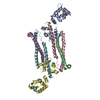
| ||||||||
|---|---|---|---|---|---|---|---|---|---|
| 1 | 
| ||||||||
| 2 | 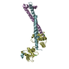
| ||||||||
| Unit cell |
|
-Components
| #1: Protein | Mass: 18673.635 Da / Num. of mol.: 2 Source method: isolated from a genetically manipulated source Source: (gene. exp.) Homo sapiens (human) / Gene: TNNC1, TNNC / Production host:  #2: Protein | Mass: 13100.999 Da / Num. of mol.: 2 Source method: isolated from a genetically manipulated source Source: (gene. exp.) Homo sapiens (human) / Gene: TNNT2 / Production host: #3: Protein | Mass: 15405.877 Da / Num. of mol.: 2 Source method: isolated from a genetically manipulated source Source: (gene. exp.) Homo sapiens (human) / Gene: TNNI3, TNNC1 / Production host: #4: Chemical | ChemComp-CA /   Mass: 40.078 Da / Num. of mol.: 6 / Source method: obtained synthetically / Formula: Ca / Feature type: SUBJECT OF INVESTIGATION Mass: 40.078 Da / Num. of mol.: 6 / Source method: obtained synthetically / Formula: Ca / Feature type: SUBJECT OF INVESTIGATIONHas ligand of interest | Y | |
|---|
-Experimental details
-Experiment
| Experiment | Method: X-RAY DIFFRACTION / Number of used crystals: 1 |
|---|
- Sample preparation
| Crystal | Density Matthews: 2.56 Å3/Da / Density % sol: 51.98 % |
|---|---|
| Crystal grow | Temperature: 291 K / Method: vapor diffusion, hanging drop / Details: 0.2M sodium acetate, 20% PEG 3350 |
-Data collection
| Diffraction | Mean temperature: 100 K / Serial crystal experiment: N |
|---|---|
| Diffraction source | Source: SYNCHROTRON / Site: APS / Beamline: 19-ID / Wavelength: 0.97973 Å |
| Detector | Type: DECTRIS PILATUS 6M / Detector: PIXEL / Date: Dec 13, 2019 |
| Radiation | Protocol: SINGLE WAVELENGTH / Monochromatic (M) / Laue (L): M / Scattering type: x-ray |
| Radiation wavelength | Wavelength: 0.97973 Å / Relative weight: 1 |
| Reflection | Resolution: 3.11→50 Å / Num. obs: 16901 / % possible obs: 99.1 % / Redundancy: 7.7 % / Rmerge(I) obs: 0.2 / Net I/σ(I): 12.6 |
| Reflection shell | Resolution: 3.11→3.26 Å / Rmerge(I) obs: 1.8 / Mean I/σ(I) obs: 1.94 / Num. unique obs: 1695 / CC1/2: 0.483 |
- Processing
| Software |
| ||||||||||||||||||||||||||||||||||||
|---|---|---|---|---|---|---|---|---|---|---|---|---|---|---|---|---|---|---|---|---|---|---|---|---|---|---|---|---|---|---|---|---|---|---|---|---|---|
| Refinement | Method to determine structure: MOLECULAR REPLACEMENT / Resolution: 3.112→41.241 Å / SU ML: 0.39 / Cross valid method: THROUGHOUT / σ(F): 1.38 / Phase error: 27.57 / Stereochemistry target values: ML
| ||||||||||||||||||||||||||||||||||||
| Solvent computation | Shrinkage radii: 0.9 Å / VDW probe radii: 1.11 Å / Solvent model: FLAT BULK SOLVENT MODEL | ||||||||||||||||||||||||||||||||||||
| Displacement parameters | Biso max: 119.98 Å2 / Biso mean: 41.8571 Å2 / Biso min: 0.09 Å2 | ||||||||||||||||||||||||||||||||||||
| Refinement step | Cycle: final / Resolution: 3.112→41.241 Å
| ||||||||||||||||||||||||||||||||||||
| LS refinement shell | Refine-ID: X-RAY DIFFRACTION / Rfactor Rfree error: 0
|
