[English] 日本語
Yorodumi
- PDB-8flv: Bruton's tyrosine kinase in complex with compound 34 -

+
Open data


ID or keywords:

Loading...

-
Basic information

Entry
Database: PDB / ID: 8flv
TitleBruton's tyrosine kinase in complex with compound 34
ComponentsTyrosine-protein kinase BTK
KeywordsTRANSFERASE/INHIBITOR / tyrosine protein kinase BTK / LIGASE / TRANSFERASE-INHIBITOR complex
Function / homology
Function and homology information


regulation of B cell cytokine production / proteoglycan catabolic process / monocyte proliferation / positive regulation of interleukin-17A production / regulation of B cell apoptotic process / eosinophil homeostasis / positive regulation of type III hypersensitivity / B cell affinity maturation / positive regulation of synoviocyte proliferation / histamine secretion by mast cell ...regulation of B cell cytokine production / proteoglycan catabolic process / monocyte proliferation / positive regulation of interleukin-17A production / regulation of B cell apoptotic process / eosinophil homeostasis / positive regulation of type III hypersensitivity / B cell affinity maturation / positive regulation of synoviocyte proliferation / histamine secretion by mast cell / neutrophil homeostasis / cellular response to molecule of fungal origin / positive regulation of type I hypersensitivity / cellular response to interleukin-7 / MyD88 deficiency (TLR2/4) / MyD88-dependent toll-like receptor signaling pathway / IRAK4 deficiency (TLR2/4) / positive regulation of immunoglobulin production / MyD88:MAL(TIRAP) cascade initiated on plasma membrane / positive regulation of B cell differentiation / positive regulation of NLRP3 inflammasome complex assembly / phospholipase activator activity / negative regulation of interleukin-10 production / B cell activation / negative regulation of B cell proliferation / Fc-epsilon receptor signaling pathway / mesoderm development / phosphatidylinositol-3,4,5-trisphosphate binding / phospholipase binding / RHO GTPases Activate WASPs and WAVEs / positive regulation of phagocytosis / positive regulation of B cell proliferation / cell maturation / FCERI mediated Ca+2 mobilization / Antigen activates B Cell Receptor (BCR) leading to generation of second messengers / calcium-mediated signaling / apoptotic signaling pathway / FCGR3A-mediated phagocytosis / B cell receptor signaling pathway / non-specific protein-tyrosine kinase / non-membrane spanning protein tyrosine kinase activity / Regulation of actin dynamics for phagocytic cup formation / G beta:gamma signalling through BTK / cellular response to reactive oxygen species / peptidyl-tyrosine phosphorylation / positive regulation of interleukin-6 production / positive regulation of tumor necrosis factor production / G alpha (12/13) signalling events / DAP12 signaling / positive regulation of NF-kappaB transcription factor activity / T cell receptor signaling pathway / ER-Phagosome pathway / cytoplasmic vesicle / G alpha (q) signalling events / protein tyrosine kinase activity / adaptive immune response / Potential therapeutics for SARS / response to lipopolysaccharide / intracellular signal transduction / membrane raft / protein phosphorylation / innate immune response / perinuclear region of cytoplasm / ATP binding / identical protein binding / nucleus / metal ion binding / plasma membrane / cytoplasm / cytosol
Similarity search - Function
Tyrosine-protein kinase BTK, SH3 domain / Zinc finger, Btk motif / BTK motif / Zinc finger Btk-type profile. / Bruton's tyrosine kinase Cys-rich motif / PH domain / PH domain profile. / Pleckstrin homology domain. / Pleckstrin homology domain / SH3 domain ...Tyrosine-protein kinase BTK, SH3 domain / Zinc finger, Btk motif / BTK motif / Zinc finger Btk-type profile. / Bruton's tyrosine kinase Cys-rich motif / PH domain / PH domain profile. / Pleckstrin homology domain. / Pleckstrin homology domain / SH3 domain / SH2 domain / Src homology 2 (SH2) domain profile. / Src homology 2 domains / SH2 domain / Src homology 3 domains / SH2 domain superfamily / SH3-like domain superfamily / Src homology 3 (SH3) domain profile. / SH3 domain / Tyrosine-protein kinase, catalytic domain / Tyrosine kinase, catalytic domain / Tyrosine protein kinases specific active-site signature. / PH-like domain superfamily / Tyrosine-protein kinase, active site / Protein tyrosine and serine/threonine kinase / Serine-threonine/tyrosine-protein kinase, catalytic domain / Protein kinase, ATP binding site / Protein kinases ATP-binding region signature. / Protein kinase domain profile. / Protein kinase domain / Protein kinase-like domain superfamily
Similarity search - Domain/homology
DI(HYDROXYETHYL)ETHER / Chem-ZB9 / Tyrosine-protein kinase BTK
Similarity search - Component
Biological speciesHomo sapiens (human)
MethodX-RAY DIFFRACTION / MOLECULAR REPLACEMENT / Resolution: 1.3 Å
AuthorsMetrick, C.M. / Marcotte, D.J.
Funding support1items
OrganizationGrant numberCountry
Not funded
CitationJournal: Bioorg.Med.Chem.Lett. / Year: 2023
Title: Discovery of structural diverse reversible BTK inhibitors utilized to develop a novel in vivo CD69 and CD86 PK/PD mouse model.
Authors: Vandeveer, G.H. / Arduini, R.M. / Baker, D.P. / Barry, K. / Bohnert, T. / Bowden-Verhoek, J.K. / Conlon, P. / Cullen, P.F. / Guan, B. / Jenkins, T.J. / Liao, S.Y. / Lin, L. / Liu, Y.T. / ...Authors: Vandeveer, G.H. / Arduini, R.M. / Baker, D.P. / Barry, K. / Bohnert, T. / Bowden-Verhoek, J.K. / Conlon, P. / Cullen, P.F. / Guan, B. / Jenkins, T.J. / Liao, S.Y. / Lin, L. / Liu, Y.T. / Marcotte, D. / Mertsching, E. / Metrick, C.M. / Negrou, E. / Powell, N. / Scott, D. / Silvian, L.F. / Hopkins, B.T.
History
DepositionDec 22, 2022Deposition site: RCSB / Processing site: RCSB
Revision 1.0Apr 5, 2023Provider: repository / Type: Initial release
Revision 1.1May 22, 2024Group: Data collection / Category: chem_comp_atom / chem_comp_bond

-
Structure visualization

Structure viewerMolecule:
MolmilJmol/JSmol

Downloads & links

-
Assembly

Deposited unit
A: Tyrosine-protein kinase BTK
hetero molecules


Theoretical massNumber of molelcules
Total (without water)33,4546
Polymers32,6801
Non-polymers7745
Water4,396244
1


  • Idetical with deposited unit
  • defined by author
  • Evidence: surface plasmon resonance
TypeNameSymmetry operationNumber
identity operation1_555x,y,z1
Unit cell
Length a, b, c (Å)71.444, 104.846, 37.964
Angle α, β, γ (deg.)90.000, 90.000, 90.000
Int Tables number18
Space group name H-MP21212
Space group name HallP22ab
Symmetry operation#1: x,y,z
#2: x+1/2,-y+1/2,-z
#3: -x+1/2,y+1/2,-z
#4: -x,-y,z

-
Components

#1: Protein Tyrosine-protein kinase BTK / Agammaglobulinemia tyrosine kinase / ATK / B-cell progenitor kinase / BPK / Bruton tyrosine kinase


Mass: 32680.471 Da / Num. of mol.: 1 / Fragment: Protein kinase domain residues 382-659
Source method: isolated from a genetically manipulated source
Source: (gene. exp.) Homo sapiens (human) / Gene: BTK, AGMX1, ATK, BPK / Production host: 
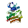
Spodoptera frugiperda (fall armyworm)
References: UniProt: Q06187, non-specific protein-tyrosine kinase
#2: Chemical ChemComp-ZB9 / 2-(3,5-dichloroanilino)-1-{(3R)-3-[methyl(7H-pyrrolo[2,3-d]pyrimidin-4-yl)amino]piperidin-1-yl}ethan-1-one


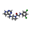
Mass: 433.334 Da / Num. of mol.: 1 / Source method: obtained synthetically / Formula: C20H22Cl2N6O / Feature type: SUBJECT OF INVESTIGATION
#3: Chemical ChemComp-DMS / DIMETHYL SULFOXIDE


Mass: 78.133 Da / Num. of mol.: 3 / Source method: obtained synthetically / Formula: C2H6OS / Comment: DMSO, precipitant*YM
#4: Chemical ChemComp-PEG / DI(HYDROXYETHYL)ETHER


Mass: 106.120 Da / Num. of mol.: 1 / Source method: obtained synthetically / Formula: C4H10O3
#5: Water ChemComp-HOH / water


Mass: 18.015 Da / Num. of mol.: 244 / Source method: isolated from a natural source / Formula: H2O
Has ligand of interestY

-
Experimental details

-
Experiment

ExperimentMethod: X-RAY DIFFRACTION / Number of used crystals: 1

-
Sample preparation

CrystalDensity Matthews: 2.28 Å3/Da / Density % sol: 46.01 %
Crystal growTemperature: 277 K / Method: vapor diffusion / Details: bis-tris, ammonium salt, PEG

-
Data collection

DiffractionMean temperature: 100 K / Serial crystal experiment: N
Diffraction sourceSource: ROTATING ANODE / Type: RIGAKU FR-E+ SUPERBRIGHT / Wavelength: 1.54 Å
DetectorType: RIGAKU RAXIS IV++ / Detector: IMAGE PLATE / Date: Aug 9, 2011
RadiationProtocol: SINGLE WAVELENGTH / Monochromatic (M) / Laue (L): M / Scattering type: x-ray
Radiation wavelengthWavelength: 1.54 Å / Relative weight: 1
ReflectionResolution: 1.3→50 Å / Num. obs: 58604 / % possible obs: 80.6 % / Redundancy: 5.4 % / Biso Wilson estimate: 12.97 Å2 / Rmerge(I) obs: 0.123 / Net I/σ(I): 6.2
Reflection shellResolution: 1.3→1.36 Å / Rmerge(I) obs: 1.687 / Num. unique obs: 2179

-
Processing

SoftwareName: PHENIX / Version: 1.20.1_4487 / Classification: refinement
RefinementMethod to determine structure: MOLECULAR REPLACEMENT / Resolution: 1.3→29.52 Å / SU ML: 0.1538 / Cross valid method: FREE R-VALUE / σ(F): 1.33 / Phase error: 24.2296
Stereochemistry target values: GeoStd + Monomer Library + CDL v1.2
RfactorNum. reflection% reflection
Rfree0.2181 2965 5.13 %
Rwork0.1941 54810 -
obs0.1954 57775 80.6 %
Solvent computationShrinkage radii: 0.9 Å / VDW probe radii: 1.1 Å / Solvent model: FLAT BULK SOLVENT MODEL
Displacement parametersBiso mean: 17.64 Å2
Refinement stepCycle: LAST / Resolution: 1.3→29.52 Å
ProteinNucleic acidLigandSolventTotal
Num. atoms2116 0 48 244 2408
Refine LS restraints
Refine-IDTypeDev idealNumber
X-RAY DIFFRACTIONf_bond_d0.00672292
X-RAY DIFFRACTIONf_angle_d0.91023108
X-RAY DIFFRACTIONf_chiral_restr0.0891329
X-RAY DIFFRACTIONf_plane_restr0.009394
X-RAY DIFFRACTIONf_dihedral_angle_d14.1196851
LS refinement shell
Resolution (Å)Rfactor RfreeNum. reflection RfreeRfactor RworkNum. reflection RworkRefine-ID% reflection obs (%)
1.3-1.320.68660.6268128X-RAY DIFFRACTION4.03
1.32-1.340.5501230.498448X-RAY DIFFRACTION14.2
1.34-1.360.449570.41641109X-RAY DIFFRACTION34.26
1.36-1.390.4302920.38641548X-RAY DIFFRACTION48.91
1.39-1.420.3274990.35041952X-RAY DIFFRACTION60.34
1.42-1.450.3731260.32972295X-RAY DIFFRACTION71.93
1.45-1.480.37371280.31132519X-RAY DIFFRACTION79.2
1.48-1.520.30661640.27822810X-RAY DIFFRACTION86.76
1.52-1.560.30361690.26362945X-RAY DIFFRACTION92.87
1.56-1.610.24821660.24473165X-RAY DIFFRACTION98.46
1.61-1.660.24011760.22143223X-RAY DIFFRACTION99.62
1.66-1.720.24341700.20773213X-RAY DIFFRACTION99.47
1.72-1.790.22641570.20283195X-RAY DIFFRACTION99.5
1.79-1.870.23441620.19263227X-RAY DIFFRACTION99.38
1.87-1.970.22271680.17573212X-RAY DIFFRACTION99.59
1.97-2.090.18781670.16733256X-RAY DIFFRACTION99.77
2.09-2.250.17961880.16323235X-RAY DIFFRACTION99.83
2.25-2.480.20641760.1713273X-RAY DIFFRACTION100
2.48-2.840.20071850.17753284X-RAY DIFFRACTION100
2.84-3.570.20692050.17533307X-RAY DIFFRACTION100
3.57-29.520.1751810.16763466X-RAY DIFFRACTION99.16

+
About Yorodumi

-
News

-
Feb 9, 2022. New format data for meta-information of EMDB entries

New format data for meta-information of EMDB entries

  • Version 3 of the EMDB header file is now the official format.
  • The previous official version 1.9 will be removed from the archive.

Related info.:EMDB header

External links:wwPDB to switch to version 3 of the EMDB data model

-
Aug 12, 2020. Covid-19 info

Covid-19 info

URL: https://pdbjlvh1.pdbj.org/emnavi/covid19.php

New page: Covid-19 featured information page in EM Navigator.

Related info.:Covid-19 info / Mar 5, 2020. Novel coronavirus structure data

+
Mar 5, 2020. Novel coronavirus structure data

Novel coronavirus structure data

Related info.:Yorodumi Speices / Aug 12, 2020. Covid-19 info

External links:COVID-19 featured content - PDBj / Molecule of the Month (242):Coronavirus Proteases

+
Jan 31, 2019. EMDB accession codes are about to change! (news from PDBe EMDB page)

EMDB accession codes are about to change! (news from PDBe EMDB page)

  • The allocation of 4 digits for EMDB accession codes will soon come to an end. Whilst these codes will remain in use, new EMDB accession codes will include an additional digit and will expand incrementally as the available range of codes is exhausted. The current 4-digit format prefixed with “EMD-” (i.e. EMD-XXXX) will advance to a 5-digit format (i.e. EMD-XXXXX), and so on. It is currently estimated that the 4-digit codes will be depleted around Spring 2019, at which point the 5-digit format will come into force.
  • The EM Navigator/Yorodumi systems omit the EMD- prefix.

Related info.:Q: What is EMD? / ID/Accession-code notation in Yorodumi/EM Navigator

External links:EMDB Accession Codes are Changing Soon! / Contact to PDBj

+
Jul 12, 2017. Major update of PDB

Major update of PDB

  • wwPDB released updated PDB data conforming to the new PDBx/mmCIF dictionary.
  • This is a major update changing the version number from 4 to 5, and with Remediation, in which all the entries are updated.
  • In this update, many items about electron microscopy experimental information are reorganized (e.g. em_software).
  • Now, EM Navigator and Yorodumi are based on the updated data.

External links:wwPDB Remediation / Enriched Model Files Conforming to OneDep Data Standards Now Available in the PDB FTP Archive

-
Yorodumi

Thousand views of thousand structures

  • Yorodumi is a browser for structure data from EMDB, PDB, SASBDB, etc.
  • This page is also the successor to EM Navigator detail page, and also detail information page/front-end page for Omokage search.
  • The word "yorodu" (or yorozu) is an old Japanese word meaning "ten thousand". "mi" (miru) is to see.

Related info.:EMDB / PDB / SASBDB / Comparison of 3 databanks / Yorodumi Search / Aug 31, 2016. New EM Navigator & Yorodumi / Yorodumi Papers / Jmol/JSmol / Function and homology information / Changes in new EM Navigator and Yorodumi

Read more