Movie
Movie Controller

[English] 日本語
 Yorodumi
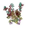
- PDB-8fk5: Cryo-EM Structure of PG9RSH DU011 Fab in complex with BG505 DS-SO... -
+ Open data

- Basic information
| Entry | Database: PDB / ID: 8fk5 | |||||||||
|---|---|---|---|---|---|---|---|---|---|---|
| Title | Cryo-EM Structure of PG9RSH DU011 Fab in complex with BG505 DS-SOSIP.664 | |||||||||
 Components Components |
| |||||||||
 Keywords Keywords | VIRAL PROTEIN / CD4 / HIV-1 / SOSIP / Vaccine / IMMUNE SYSTEM / llama / V2 apex / therapeutic | |||||||||
| Function / homology |  Function and homology information Function and homology informationimmunoglobulin complex / symbiont-mediated perturbation of host defense response / positive regulation of plasma membrane raft polarization / positive regulation of receptor clustering / host cell endosome membrane / clathrin-dependent endocytosis of virus by host cell / adaptive immune response / viral protein processing / fusion of virus membrane with host plasma membrane / fusion of virus membrane with host endosome membrane ...immunoglobulin complex / symbiont-mediated perturbation of host defense response / positive regulation of plasma membrane raft polarization / positive regulation of receptor clustering / host cell endosome membrane / clathrin-dependent endocytosis of virus by host cell / adaptive immune response / viral protein processing / fusion of virus membrane with host plasma membrane / fusion of virus membrane with host endosome membrane / viral envelope / virion attachment to host cell / host cell plasma membrane / virion membrane / structural molecule activity / extracellular region / membrane / identical protein binding / plasma membrane Similarity search - Function | |||||||||
| Biological species |   Human immunodeficiency virus 1 Human immunodeficiency virus 1 Homo sapiens (human) Homo sapiens (human) | |||||||||
| Method | ELECTRON MICROSCOPY / single particle reconstruction / cryo EM / Resolution: 3.4 Å | |||||||||
 Authors Authors | Gorman, J. / Kwong, P.D. | |||||||||
| Funding support |  United States, 2items United States, 2items
| |||||||||
 Citation Citation | Journal: Cell Rep / Year: 2023 Title: Improved HIV-1 neutralization breadth and potency of V2-apex antibodies by in silico design. Authors: Graham T Holt / Jason Gorman / Siyu Wang / Anna U Lowegard / Baoshan Zhang / Tracy Liu / Bob C Lin / Mark K Louder / Marcel S Frenkel / Krisha McKee / Sijy O'Dell / Reda Rawi / Chen-Hsiang ...Authors: Graham T Holt / Jason Gorman / Siyu Wang / Anna U Lowegard / Baoshan Zhang / Tracy Liu / Bob C Lin / Mark K Louder / Marcel S Frenkel / Krisha McKee / Sijy O'Dell / Reda Rawi / Chen-Hsiang Shen / Nicole A Doria-Rose / Peter D Kwong / Bruce R Donald / Abstract: Broadly neutralizing antibodies (bNAbs) against HIV can reduce viral transmission in humans, but an effective therapeutic will require unusually high breadth and potency of neutralization. We employ ...Broadly neutralizing antibodies (bNAbs) against HIV can reduce viral transmission in humans, but an effective therapeutic will require unusually high breadth and potency of neutralization. We employ the OSPREY computational protein design software to engineer variants of two apex-directed bNAbs, PGT145 and PG9RSH, resulting in increases in potency of over 100-fold against some viruses. The top designed variants improve neutralization breadth from 39% to 54% at clinically relevant concentrations (IC < 1 μg/mL) and improve median potency (IC) by up to 4-fold over a cross-clade panel of 208 strains. To investigate the mechanisms of improvement, we determine cryoelectron microscopy structures of each variant in complex with the HIV envelope trimer. Surprisingly, we find the largest increases in breadth to be a result of optimizing side-chain interactions with highly variable epitope residues. These results provide insight into mechanisms of neutralization breadth and inform strategies for antibody design and improvement. | |||||||||
| History |
|
- Structure visualization
| Structure viewer | Molecule: MolmilJmol/JSmol |
|---|
- Downloads & links
-Download
| PDBx/mmCIF format | 8fk5.cif.gz | 390 KB | Display | PDBx/mmCIF format |
|---|---|---|---|---|
| PDB format | pdb8fk5.ent.gz | Display | PDB format | |
| PDBx/mmJSON format | 8fk5.json.gz | Tree view | PDBx/mmJSON format | |
| Others |  Other downloads Other downloads |
-Validation report
| Arichive directory | https://data.pdbj.org/pub/pdb/validation_reports/fk/8fk5ftp://data.pdbj.org/pub/pdb/validation_reports/fk/8fk5 | HTTPS FTP |
|---|
-Related structure data
| Related structure data | 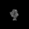 29248MC  8fl1C  8flwC M: map data used to model this data C: citing same article ( |
|---|---|
| Similar structure data |


-Links
 PDBj
PDBj


- Assembly
| Deposited unit | 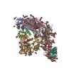
|
|---|---|
| 1 |
|
-Components
-Envelope glycoprotein ... , 2 types, 6 molecules ABFCGI
| #1: Protein | Mass: 17146.482 Da / Num. of mol.: 3 / Fragment: UNP residues 509-661 Source method: isolated from a genetically manipulated source Source: (gene. exp.) Human immunodeficiency virus 1 / Gene: env / Production host: Homo sapiens (human) / References: UniProt: Q2N0S6#2: Protein | Mass: 54086.324 Da / Num. of mol.: 3 / Fragment: UNP residues 30-510 Source method: isolated from a genetically manipulated source Source: (gene. exp.) Human immunodeficiency virus 1 / Strain: BG505 / Gene: env / Production host: Homo sapiens (human) / References: UniProt: Q2N0S6 |
|---|
-Antibody , 2 types, 2 molecules HL
| #3: Antibody | Mass: 27260.258 Da / Num. of mol.: 1 Source method: isolated from a genetically manipulated source Source: (gene. exp.) Homo sapiens (human) / Gene: VHCH1 / Production host: Homo sapiens (human) / References: UniProt: A4F255 |
|---|---|
| #4: Antibody | Mass: 22804.182 Da / Num. of mol.: 1 Source method: isolated from a genetically manipulated source Source: (gene. exp.) Homo sapiens (human) / Production host: Homo sapiens (human) / References: UniProt: Q6PJG0 |
-Sugars , 5 types, 61 molecules 
| #5: Polysaccharide | 2-acetamido-2-deoxy-beta-D-glucopyranose-(1-4)-2-acetamido-2-deoxy-beta-D-glucopyranose Source method: isolated from a genetically manipulated source #6: Polysaccharide | Source method: isolated from a genetically manipulated source #7: Polysaccharide | Source method: isolated from a genetically manipulated source #8: Polysaccharide | alpha-D-mannopyranose-(1-2)-alpha-D-mannopyranose-(1-6)-[alpha-D-mannopyranose-(1-3)]alpha-D- ...alpha-D-mannopyranose-(1-2)-alpha-D-mannopyranose-(1-6)-[alpha-D-mannopyranose-(1-3)]alpha-D-mannopyranose-(1-6)-[alpha-D-mannopyranose-(1-2)-alpha-D-mannopyranose-(1-3)]beta-D-mannopyranose-(1-4)-2-acetamido-2-deoxy-beta-D-glucopyranose-(1-4)-2-acetamido-2-deoxy-beta-D-glucopyranose | Source method: isolated from a genetically manipulated source #9: Sugar | ChemComp-NAG /  Type: D-saccharide, beta linking / Mass: 221.208 Da / Num. of mol.: 38 / Source method: obtained synthetically / Formula: C8H15NO6 Type: D-saccharide, beta linking / Mass: 221.208 Da / Num. of mol.: 38 / Source method: obtained synthetically / Formula: C8H15NO6 |
|---|
-Details
| Has ligand of interest | N |
|---|---|
| Has protein modification | Y |
-Experimental details
-Experiment
| Experiment | Method: ELECTRON MICROSCOPY |
|---|---|
| EM experiment | Aggregation state: PARTICLE / 3D reconstruction method: single particle reconstruction |
- Sample preparation
| Component | Name: Cryo-EM Structure of PG9RSH DU011 Fab in complex with BG505 DS-SOSIP.664 Type: COMPLEX / Entity ID: #1-#4 / Source: RECOMBINANT |
|---|---|
| Molecular weight | Experimental value: NO |
| Source (natural) | Organism: Human immunodeficiency virus 1 |
| Source (recombinant) | Organism: Homo sapiens (human) |
| Buffer solution | pH: 7.4 / Details: PBS |
| Specimen | Conc.: 2 mg/ml / Embedding applied: NO / Shadowing applied: NO / Staining applied: NO / Vitrification applied: YES |
| Specimen support | Grid type: C-flat-1.2/1.3 |
| Vitrification | Instrument: FEI VITROBOT MARK IV / Cryogen name: ETHANE / Humidity: 90 % / Chamber temperature: 293 K |
- Electron microscopy imaging
| Experimental equipment |  Model: Titan Krios / Image courtesy: FEI Company |
|---|---|
| Microscopy | Model: FEI TITAN KRIOS |
| Electron gun | Electron source:  FIELD EMISSION GUN / Accelerating voltage: 300 kV / Illumination mode: FLOOD BEAM FIELD EMISSION GUN / Accelerating voltage: 300 kV / Illumination mode: FLOOD BEAM |
| Electron lens | Mode: BRIGHT FIELD / Nominal defocus max: 2500 nm / Nominal defocus min: 800 nm / C2 aperture diameter: 70 µm |
| Image recording | Average exposure time: 2 sec. / Electron dose: 63.75 e/Å2 / Detector mode: COUNTING / Film or detector model: GATAN K3 (6k x 4k) / Num. of grids imaged: 1 |
- Processing
| Software | Name: PHENIX / Version: 1.19.2_4158: / Classification: refinement | ||||||||||||||||||||||||
|---|---|---|---|---|---|---|---|---|---|---|---|---|---|---|---|---|---|---|---|---|---|---|---|---|---|
| EM software |
| ||||||||||||||||||||||||
| CTF correction | Type: PHASE FLIPPING AND AMPLITUDE CORRECTION | ||||||||||||||||||||||||
| 3D reconstruction | Resolution: 3.4 Å / Resolution method: FSC 0.143 CUT-OFF / Num. of particles: 230180 / Symmetry type: POINT | ||||||||||||||||||||||||
| Atomic model building | Protocol: FLEXIBLE FIT / Space: REAL | ||||||||||||||||||||||||
| Atomic model building | PDB-ID: 6NNF Accession code: 6NNF / Source name: PDB / Type: experimental model | ||||||||||||||||||||||||
| Refine LS restraints |
|