 Movie
Movie Controller


+ Open data

- Basic information
| Entry | Database: PDB / ID: 8fjl | |||||||||||||||||||||
|---|---|---|---|---|---|---|---|---|---|---|---|---|---|---|---|---|---|---|---|---|---|---|
| Title | Golden Shiner Reovirus Core Tropical Vertex | |||||||||||||||||||||
 Components Components |
| |||||||||||||||||||||
 Keywords Keywords | VIRUS / dsRNA / RNA-dependent / RNA-polymerase | |||||||||||||||||||||
| Function / homology |  Function and homology information Function and homology informationviral intermediate capsid / T=2 icosahedral viral capsid / host cytoskeleton / viral inner capsid / viral outer capsid / 7-methylguanosine mRNA capping / viral genome replication / viral nucleocapsid / mRNA guanylyltransferase / mRNA guanylyltransferase activity ...viral intermediate capsid / T=2 icosahedral viral capsid / host cytoskeleton / viral inner capsid / viral outer capsid / 7-methylguanosine mRNA capping / viral genome replication / viral nucleocapsid / mRNA guanylyltransferase / mRNA guanylyltransferase activity / mRNA (guanine-N7)-methyltransferase / mRNA 5'-cap (guanine-N7-)-methyltransferase activity / RNA helicase activity / RNA-directed RNA polymerase / nucleotide binding / hydrolase activity / RNA-directed RNA polymerase activity / GTP binding / structural molecule activity / RNA binding / zinc ion binding / ATP binding Similarity search - Function | |||||||||||||||||||||
| Biological species |  Golden shiner reovirus Golden shiner reovirus | |||||||||||||||||||||
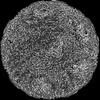
| Method | ELECTRON MICROSCOPY / single particle reconstruction / cryo EM / Resolution: 3.27 Å | |||||||||||||||||||||
 Authors Authors | Stevens, A.S. / Zhou, Z.H. | |||||||||||||||||||||
| Funding support |  United States, 6items United States, 6items
| |||||||||||||||||||||
 Citation Citation | Journal: Protein Cell / Year: 2023 Title: Asymmetric reconstruction of the aquareovirus core at near-atomic resolution and mechanism of transcription initiation. Authors: Alexander Stevens / Yanxiang Cui / Sakar Shivakoti / Z Hong Zhou / | |||||||||||||||||||||
| History |
|
- Structure visualization
| Structure viewer | Molecule: MolmilJmol/JSmol |
|---|
- Downloads & links
-Download
| PDBx/mmCIF format | 8fjl.cif.gz | 4.2 MB | Display | PDBx/mmCIF format |
|---|---|---|---|---|
| PDB format | pdb8fjl.ent.gz | Display | PDB format | |
| PDBx/mmJSON format | 8fjl.json.gz | Tree view | PDBx/mmJSON format | |
| Others |  Other downloads Other downloads |
-Validation report
| Arichive directory | https://data.pdbj.org/pub/pdb/validation_reports/fj/8fjlftp://data.pdbj.org/pub/pdb/validation_reports/fj/8fjl | HTTPS FTP |
|---|
-Related structure data
| Related structure data |  29244MC  8fjkC M: map data used to model this data C: citing same article ( |
|---|---|
| Similar structure data |


-Links
 PDBj
PDBj


- Assembly
| Deposited unit | 
|
|---|---|
| 1 |
|
-Components
-Protein , 4 types, 17 molecules ABVWXabdeghnYZcfi
| #1: Protein | Mass: 141605.281 Da / Num. of mol.: 1 / Source method: isolated from a natural source / Source: (natural) Golden shiner reovirus / References: UniProt: Q8JU61, RNA-directed RNA polymerase | ||
|---|---|---|---|
| #2: Protein | Mass: 79347.359 Da / Num. of mol.: 1 / Source method: isolated from a natural source / Source: (natural) Golden shiner reovirus / References: UniProt: Q8JU58 | ||
| #9: Protein | Mass: 44489.391 Da / Num. of mol.: 10 / Source method: isolated from a natural source / Source: (natural) Golden shiner reovirus / References: UniProt: Q8JU54#10: Protein | Mass: 141109.750 Da / Num. of mol.: 5 / Source method: isolated from a natural source / Source: (natural) Golden shiner reovirusReferences: UniProt: Q8JU62, mRNA guanylyltransferase, mRNA (guanine-N7)-methyltransferase |
-Major inner capsid protein ... , 2 types, 15 molecules CDEFGHIJKLMNklm
| #3: Protein | Mass: 124694.531 Da / Num. of mol.: 10 / Source method: isolated from a natural source / Source: (natural) Golden shiner reovirus / References: UniProt: Q8JU60, RNA helicase#4: Protein | Mass: 9147.655 Da / Num. of mol.: 5 / Fragment: N-terminal residues 13-106 / Source method: isolated from a natural source / Source: (natural) Golden shiner reovirus / References: UniProt: Q8JU60, RNA helicase |
|---|
-RNA chain , 5 types, 10 molecules a5b5b6a6a1b1a3b3a2b2
| #5: RNA chain | Mass: 8850.246 Da / Num. of mol.: 2 / Source method: isolated from a natural source / Source: (natural) Golden shiner reovirus#6: RNA chain | Mass: 12027.101 Da / Num. of mol.: 2 / Source method: isolated from a natural source / Source: (natural) Golden shiner reovirus#7: RNA chain | Mass: 16474.699 Da / Num. of mol.: 2 / Source method: isolated from a natural source / Source: (natural) Golden shiner reovirus#8: RNA chain | Mass: 12662.472 Da / Num. of mol.: 2 / Source method: isolated from a natural source / Source: (natural) Golden shiner reovirus#11: RNA chain | Mass: 19016.188 Da / Num. of mol.: 2 / Source method: isolated from a natural source / Source: (natural) Golden shiner reovirus |
|---|
-Non-polymers , 1 types, 4 molecules 
| #12: Chemical | ChemComp-ZN /  Mass: 65.409 Da / Num. of mol.: 4 / Source method: obtained synthetically / Formula: Zn Mass: 65.409 Da / Num. of mol.: 4 / Source method: obtained synthetically / Formula: Zn |
|---|
-Details
| Has ligand of interest | N |
|---|
-Experimental details
-Experiment
| Experiment | Method: ELECTRON MICROSCOPY |
|---|---|
| EM experiment | Aggregation state: PARTICLE / 3D reconstruction method: single particle reconstruction |
- Sample preparation
| Component | Name: Golden shiner reovirus / Type: VIRUS / Details: Virus cultured in fathead minnow cells / Entity ID: #1-#11 / Source: NATURAL |
|---|---|
| Molecular weight | Value: 47 MDa / Experimental value: NO |
| Source (natural) | Organism: Golden shiner reovirus |
| Details of virus | Empty: NO / Enveloped: NO / Isolate: STRAIN / Type: VIRION |
| Natural host | Organism: Notemigonus crysoleucas |
| Buffer solution | pH: 7.4 / Details: Phosphate buffered saline |
| Specimen | Embedding applied: NO / Shadowing applied: NO / Staining applied: NO / Vitrification applied: YES |
| Specimen support | Grid type: Quantifoil R2/1 |
| Vitrification | Instrument: FEI VITROBOT MARK IV / Cryogen name: ETHANE / Humidity: 100 % / Chamber temperature: 277 K |
- Electron microscopy imaging
| Experimental equipment |  Model: Titan Krios / Image courtesy: FEI Company |
|---|---|
| Microscopy | Model: FEI TITAN KRIOS |
| Electron gun | Electron source:  FIELD EMISSION GUN / Accelerating voltage: 300 kV / Illumination mode: FLOOD BEAM FIELD EMISSION GUN / Accelerating voltage: 300 kV / Illumination mode: FLOOD BEAM |
| Electron lens | Mode: BRIGHT FIELD / Nominal magnification: 81000 X / Calibrated magnification: 81000 X / Nominal defocus max: 2500 nm / Nominal defocus min: 1500 nm / Calibrated defocus min: 1500 nm / Calibrated defocus max: 2500 nm / Cs: 2.7 mm / C2 aperture diameter: 50 µm / Alignment procedure: COMA FREE |
| Specimen holder | Cryogen: NITROGEN / Specimen holder model: FEI TITAN KRIOS AUTOGRID HOLDER |
| Image recording | Electron dose: 45 e/Å2 / Detector mode: SUPER-RESOLUTION / Film or detector model: GATAN K2 SUMMIT (4k x 4k) |
- Processing
| Software | Name: UCSF ChimeraX / Version: 1.5/v9 / Classification: model building / URL: https://www.rbvi.ucsf.edu/chimerax/ / Os: Windows / Type: package | |||||||||||||||
|---|---|---|---|---|---|---|---|---|---|---|---|---|---|---|---|---|
| EM software |
| |||||||||||||||
| CTF correction | Type: PHASE FLIPPING AND AMPLITUDE CORRECTION | |||||||||||||||
| Particle selection | Num. of particles selected: 99323 | |||||||||||||||
| 3D reconstruction | Resolution: 3.27 Å / Resolution method: FSC 0.143 CUT-OFF / Num. of particles: 99323 / Symmetry type: POINT |
