 ムービー
ムービー コントローラー


+ データを開く

- 基本情報
基本情報
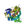
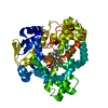
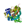
| 登録情報 | データベース: PDB / ID: 8fic | |||||||||
|---|---|---|---|---|---|---|---|---|---|---|
| タイトル | Crystal Structure of Erwinia tracheiphila CYP114 in complex with ent-kaurenoic acid (Crystal Form 1) | |||||||||
 要素 要素 | Cytochrome P450 | |||||||||
 キーワード キーワード | OXIDOREDUCTASE / gibberellin | |||||||||
| 機能・相同性 |  機能・相同性情報 機能・相同性情報oxidoreductase activity, acting on paired donors, with incorporation or reduction of molecular oxygen / monooxygenase activity / iron ion binding / heme binding 類似検索 - 分子機能 | |||||||||
| 生物種 |  Erwinia tracheiphila (バクテリア) Erwinia tracheiphila (バクテリア) | |||||||||
| 手法 |  X線回折 / シンクロトロン / 分子置換 / 解像度: 1.7 Å X線回折 / シンクロトロン / 分子置換 / 解像度: 1.7 Å | |||||||||
 データ登録者 データ登録者 | Stewart Jr., C.E. / Nagel, R. | |||||||||
| 資金援助 |  米国, 米国,  ドイツ, 2件 ドイツ, 2件
| |||||||||
 引用 引用 | ジャーナル: Proc.Natl.Acad.Sci.USA / 年: 2023 タイトル: Dual factors required for cytochrome-P450-mediated hydrocarbon ring contraction in bacterial gibberellin phytohormone biosynthesis. 著者: Nagel, R. / Alexander, L.E. / Stewart Jr., C.E. / Peters, R.J. | |||||||||
| 履歴 |
|
- 構造の表示
構造の表示
| 構造ビューア | 分子: MolmilJmol/JSmol |
|---|
- ダウンロードとリンク
-ダウンロード
| PDBx/mmCIF形式 | 8fic.cif.gz | 325.5 KB | 表示 | PDBx/mmCIF形式 |
|---|---|---|---|---|
| PDB形式 | pdb8fic.ent.gz | 221 KB | 表示 | PDB形式 |
| PDBx/mmJSON形式 | 8fic.json.gz | ツリー表示 | PDBx/mmJSON形式 | |
| その他 |  その他のダウンロード その他のダウンロード |
-検証レポート
| 文書・要旨 | 8fic_validation.pdf.gz | 1.1 MB | 表示 | wwPDB検証レポート |
|---|---|---|---|---|
| 文書・詳細版 | 8fic_full_validation.pdf.gz | 1.1 MB | 表示 | |
| XML形式データ | 8fic_validation.xml.gz | 19.3 KB | 表示 | |
| CIF形式データ | 8fic_validation.cif.gz | 28.5 KB | 表示 | |
| アーカイブディレクトリ | https://data.pdbj.org/pub/pdb/validation_reports/fi/8ficftp://data.pdbj.org/pub/pdb/validation_reports/fi/8fic | HTTPS FTP |
-関連構造データ
-リンク
 PDBj
PDBj
- 集合体
集合体
| 登録構造単位 | 
| ||||||||||||
|---|---|---|---|---|---|---|---|---|---|---|---|---|---|
| 1 |
| ||||||||||||
| 単位格子 |
|
-要素
| #1: タンパク質 | 分子量: 48881.285 Da / 分子数: 1 / 由来タイプ: 組換発現 由来: (組換発現) Erwinia tracheiphila (バクテリア)遺伝子: SY86_19565 / プラスミド: pET101 / 発現宿主: |
|---|---|
| #2: 化合物 | ChemComp-HEM /   分子量: 616.487 Da / 分子数: 1 / 由来タイプ: 合成 / 式: C34H32FeN4O4 / タイプ: SUBJECT OF INVESTIGATION 分子量: 616.487 Da / 分子数: 1 / 由来タイプ: 合成 / 式: C34H32FeN4O4 / タイプ: SUBJECT OF INVESTIGATION |
| #3: 化合物 | ChemComp-NE4 / (  分子量: 302.451 Da / 分子数: 1 / 由来タイプ: 合成 / 式: C20H30O2 / タイプ: SUBJECT OF INVESTIGATION 分子量: 302.451 Da / 分子数: 1 / 由来タイプ: 合成 / 式: C20H30O2 / タイプ: SUBJECT OF INVESTIGATION |
| #4: 水 | ChemComp-HOH /  分子量: 18.015 Da / 分子数: 269 / 由来タイプ: 天然 / 式: H2O 分子量: 18.015 Da / 分子数: 269 / 由来タイプ: 天然 / 式: H2O |
| 研究の焦点であるリガンドがあるか | Y |
-実験情報
-実験
| 実験 | 手法: X線回折 / 使用した結晶の数: 1 |
|---|
- 試料調製
試料調製
| 結晶 | マシュー密度: 2.33 Å3/Da / 溶媒含有率: 47.27 % / Mosaicity: 0.32 ° |
|---|---|
| 結晶化 | 温度: 291 K / 手法: 蒸気拡散法 / pH: 5.6 詳細: 16% PEG3350, 0.1M MES/Tris pH5.6, 0.2M Magnesium chloride hexahydrate |
-データ収集
| 回折 | 平均測定温度: 100 K / Serial crystal experiment: N | ||||||||||||||||||||||||||||||
|---|---|---|---|---|---|---|---|---|---|---|---|---|---|---|---|---|---|---|---|---|---|---|---|---|---|---|---|---|---|---|---|
| 放射光源 | 由来: シンクロトロン / サイト: APS / ビームライン: 23-ID-D / 波長: 1.0332 Å | ||||||||||||||||||||||||||||||
| 検出器 | タイプ: DECTRIS PILATUS3 6M / 検出器: PIXEL / 日付: 2018年6月10日 / 詳細: mirrors | ||||||||||||||||||||||||||||||
| 放射 | モノクロメーター: Si(111) / プロトコル: SINGLE WAVELENGTH / 単色(M)・ラウエ(L): M / 散乱光タイプ: x-ray | ||||||||||||||||||||||||||||||
| 放射波長 | 波長: 1.0332 Å / 相対比: 1 | ||||||||||||||||||||||||||||||
| 反射 | 解像度: 1.7→70.46 Å / Num. obs: 51325 / % possible obs: 99.9 % / 冗長度: 7.8 % / CC1/2: 0.994 / Rmerge(I) obs: 0.131 / Rpim(I) all: 0.048 / Rrim(I) all: 0.141 / Net I/σ(I): 7.5 / Num. measured all: 399178 / Scaling rejects: 142 | ||||||||||||||||||||||||||||||
| 反射 シェル | Diffraction-ID: 1
|
-位相決定
| 位相決定 | 手法: 分子置換 | |||||||||
|---|---|---|---|---|---|---|---|---|---|---|
| Phasing MR |
|
- 解析
解析
| ソフトウェア |
| |||||||||||||||||||||||||||||||||||||||||||||||||||||||||||||||||||||||||||||||||||||||||||||||||||||||||
|---|---|---|---|---|---|---|---|---|---|---|---|---|---|---|---|---|---|---|---|---|---|---|---|---|---|---|---|---|---|---|---|---|---|---|---|---|---|---|---|---|---|---|---|---|---|---|---|---|---|---|---|---|---|---|---|---|---|---|---|---|---|---|---|---|---|---|---|---|---|---|---|---|---|---|---|---|---|---|---|---|---|---|---|---|---|---|---|---|---|---|---|---|---|---|---|---|---|---|---|---|---|---|---|---|---|---|
| 精密化 | 構造決定の手法: 分子置換 開始モデル: 7RLP  7rlp 解像度: 1.7→59.34 Å / SU ML: 0.2119 / 交差検証法: FREE R-VALUE / σ(F): 1.34 / 位相誤差: 19.6455 立体化学のターゲット値: GeoStd + Monomer Library + CDL v1.2
| |||||||||||||||||||||||||||||||||||||||||||||||||||||||||||||||||||||||||||||||||||||||||||||||||||||||||
| 溶媒の処理 | 減衰半径: 0.9 Å / VDWプローブ半径: 1.11 Å / 溶媒モデル: FLAT BULK SOLVENT MODEL | |||||||||||||||||||||||||||||||||||||||||||||||||||||||||||||||||||||||||||||||||||||||||||||||||||||||||
| 原子変位パラメータ | Biso mean: 39.43 Å2 | |||||||||||||||||||||||||||||||||||||||||||||||||||||||||||||||||||||||||||||||||||||||||||||||||||||||||
| 精密化ステップ | サイクル: LAST / 解像度: 1.7→59.34 Å
| |||||||||||||||||||||||||||||||||||||||||||||||||||||||||||||||||||||||||||||||||||||||||||||||||||||||||
| 拘束条件 |
| |||||||||||||||||||||||||||||||||||||||||||||||||||||||||||||||||||||||||||||||||||||||||||||||||||||||||
| LS精密化 シェル |
|
