 Movie
Movie Controller

[English] 日本語
 Yorodumi
- PDB-8fi4: Crystal Structure of Glycylpeptide N-tetradecanoyltransferase (N-... -
+ Open data

- Basic information
| Entry | Database: PDB / ID: 8fi4 | |||||||||
|---|---|---|---|---|---|---|---|---|---|---|
| Title | Crystal Structure of Glycylpeptide N-tetradecanoyltransferase (N-myristoyl transferase) (NMT) from Leishmania major Friedlin bound to tetradecanoyl-CoA (Orthorhombic P Form 1) | |||||||||
 Components Components | Glycylpeptide N-tetradecanoyltransferase | |||||||||
 Keywords Keywords | TRANSFERASE / SSGCID / STRUCTURAL GENOMICS / SEATTLE STRUCTURAL GENOMICS CENTER FOR INFECTIOUS DISEASE | |||||||||
| Function / homology | TETRADECANOYL-COA / :  Function and homology information Function and homology information | |||||||||
| Biological species |  Leishmania major strain Friedlin (eukaryote) Leishmania major strain Friedlin (eukaryote) | |||||||||
| Method |  X-RAY DIFFRACTION / SYNCHROTRON / MOLECULAR REPLACEMENT / Resolution: 1.8 Å X-RAY DIFFRACTION / SYNCHROTRON / MOLECULAR REPLACEMENT / Resolution: 1.8 Å | |||||||||
 Authors Authors | Seattle Structural Genomics Center for Infectious Disease / Seattle Structural Genomics Center for Infectious Disease (SSGCID) | |||||||||
| Funding support |  United States, 2items United States, 2items
| |||||||||
 Citation Citation | Journal: To be published Title: Crystal Structure of Glycylpeptide N-tetradecanoyltransferase (N-myristoyl transferase) (NMT) from Leishmania major Friedlin bound to tetradecanoyl-CoA (Orthorhombic P Form 1) Authors: Lovell, S. / Battaile, K.P. / Staker, B.L. / Liu, L. | |||||||||
| History |
|
- Structure visualization
| Structure viewer | Molecule: MolmilJmol/JSmol |
|---|
- Downloads & links
-Download
| PDBx/mmCIF format | 8fi4.cif.gz | 105.8 KB | Display | PDBx/mmCIF format |
|---|---|---|---|---|
| PDB format | pdb8fi4.ent.gz | 76.3 KB | Display | PDB format |
| PDBx/mmJSON format | 8fi4.json.gz | Tree view | PDBx/mmJSON format | |
| Others |  Other downloads Other downloads |
-Validation report
| Arichive directory | https://data.pdbj.org/pub/pdb/validation_reports/fi/8fi4ftp://data.pdbj.org/pub/pdb/validation_reports/fi/8fi4 | HTTPS FTP |
|---|
-Related structure data
| Similar structure data |
|---|
-Links
 PDBj
PDBj- Assembly
| Deposited unit | 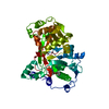
| ||||||||
|---|---|---|---|---|---|---|---|---|---|
| 1 |
| ||||||||
| Unit cell |
|
-Components
| #1: Protein | Mass: 49174.914 Da / Num. of mol.: 1 / Mutation: K148R Source method: isolated from a genetically manipulated source Source: (gene. exp.) Leishmania major strain Friedlin (eukaryote)Gene: LMJFC_320006100 / Plasmid: LemaA.18219.a.B2 / Production host:  |
|---|---|
| #2: Chemical | ChemComp-MYA /   Mass: 977.890 Da / Num. of mol.: 1 / Source method: obtained synthetically / Formula: C35H62N7O17P3S / Feature type: SUBJECT OF INVESTIGATION Mass: 977.890 Da / Num. of mol.: 1 / Source method: obtained synthetically / Formula: C35H62N7O17P3S / Feature type: SUBJECT OF INVESTIGATION |
| #3: Water | ChemComp-HOH /  Mass: 18.015 Da / Num. of mol.: 203 / Source method: isolated from a natural source / Formula: H2O Mass: 18.015 Da / Num. of mol.: 203 / Source method: isolated from a natural source / Formula: H2O |
| Has ligand of interest | Y |
-Experimental details
-Experiment
| Experiment | Method: X-RAY DIFFRACTION / Number of used crystals: 1 |
|---|
- Sample preparation
| Crystal | Density Matthews: 1.99 Å3/Da / Density % sol: 38.26 % |
|---|---|
| Crystal grow | Temperature: 291 K / Method: vapor diffusion, sitting drop / pH: 9 Details: 0.1 M Bicine, 20% PEG 6000, LemaA.18219.a.B2.PS38707 at 11.17 mg/mL. Tray: Tray 383B3/1 mM Perez-1, Puck: HR00406_02, Cryo: 20% PEG 200. MYA ligand was acquired from the expression host and ...Details: 0.1 M Bicine, 20% PEG 6000, LemaA.18219.a.B2.PS38707 at 11.17 mg/mL. Tray: Tray 383B3/1 mM Perez-1, Puck: HR00406_02, Cryo: 20% PEG 200. MYA ligand was acquired from the expression host and not added prior to crystallization. |
-Data collection
| Diffraction | Mean temperature: 100 K / Serial crystal experiment: N |
|---|---|
| Diffraction source | Source: SYNCHROTRON / Site: NSLS-II / Beamline: 19-ID / Wavelength: 0.9786 Å |
| Detector | Type: DECTRIS EIGER2 XE 9M / Detector: PIXEL / Date: Dec 10, 2022 |
| Radiation | Monochromator: Double Crystal Si 111 / Protocol: SINGLE WAVELENGTH / Monochromatic (M) / Laue (L): M / Scattering type: x-ray |
| Radiation wavelength | Wavelength: 0.9786 Å / Relative weight: 1 |
| Reflection | Resolution: 1.8→46.39 Å / Num. obs: 36370 / % possible obs: 98.4 % / Redundancy: 10.7 % / CC1/2: 0.998 / Rmerge(I) obs: 0.122 / Rpim(I) all: 0.039 / Rrim(I) all: 0.128 / Χ2: 0.95 / Net I/σ(I): 12.2 / Num. measured all: 387821 |
| Reflection shell | Resolution: 1.8→1.84 Å / % possible obs: 97.2 % / Redundancy: 11.5 % / Rmerge(I) obs: 1.388 / Num. measured all: 23955 / Num. unique obs: 2090 / CC1/2: 0.803 / Rpim(I) all: 0.423 / Rrim(I) all: 1.452 / Χ2: 0.87 / Net I/σ(I) obs: 2 |
- Processing
| Software |
| ||||||||||||||||||||||||||||||||||||||||||||||||||||||||||||||||||||||||||||||||||||||||||||||||||
|---|---|---|---|---|---|---|---|---|---|---|---|---|---|---|---|---|---|---|---|---|---|---|---|---|---|---|---|---|---|---|---|---|---|---|---|---|---|---|---|---|---|---|---|---|---|---|---|---|---|---|---|---|---|---|---|---|---|---|---|---|---|---|---|---|---|---|---|---|---|---|---|---|---|---|---|---|---|---|---|---|---|---|---|---|---|---|---|---|---|---|---|---|---|---|---|---|---|---|---|
| Refinement | Method to determine structure: MOLECULAR REPLACEMENT / Resolution: 1.8→46.39 Å / SU ML: 0.17 / Cross valid method: FREE R-VALUE / σ(F): 1.35 / Phase error: 18.65 / Stereochemistry target values: ML
| ||||||||||||||||||||||||||||||||||||||||||||||||||||||||||||||||||||||||||||||||||||||||||||||||||
| Solvent computation | Shrinkage radii: 0.9 Å / VDW probe radii: 1.1 Å / Solvent model: FLAT BULK SOLVENT MODEL | ||||||||||||||||||||||||||||||||||||||||||||||||||||||||||||||||||||||||||||||||||||||||||||||||||
| Refinement step | Cycle: LAST / Resolution: 1.8→46.39 Å
| ||||||||||||||||||||||||||||||||||||||||||||||||||||||||||||||||||||||||||||||||||||||||||||||||||
| Refine LS restraints |
| ||||||||||||||||||||||||||||||||||||||||||||||||||||||||||||||||||||||||||||||||||||||||||||||||||
| LS refinement shell |
|
