 Movie
Movie Controller

[English] 日本語
 Yorodumi
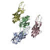
- PDB-8fha: Intramolecular ester bond-containing repeat domain 1 from Suiprao... -
+ Open data

- Basic information
| Entry | Database: PDB / ID: 8fha | ||||||
|---|---|---|---|---|---|---|---|
| Title | Intramolecular ester bond-containing repeat domain 1 from Suipraoptans intestinalis adhesin | ||||||
 Components Components | Adhesin | ||||||
 Keywords Keywords | CELL ADHESION / bacterial adhesin domain / Ig-like domain / intramolecular ester bond | ||||||
| Function / homology |  Function and homology information Function and homology information | ||||||
| Biological species |  Suipraeoptans intestinalis (bacteria) Suipraeoptans intestinalis (bacteria) | ||||||
| Method |  X-RAY DIFFRACTION / SYNCHROTRON / MOLECULAR REPLACEMENT / Resolution: 1.49 Å X-RAY DIFFRACTION / SYNCHROTRON / MOLECULAR REPLACEMENT / Resolution: 1.49 Å | ||||||
 Authors Authors | Jin, S. / Young, P.G. / Squire, C.J. | ||||||
| Funding support | 1items
| ||||||
 Citation Citation | Journal: To Be Published Title: Intramolecular ester bond-containing repeat domain from Suipraoptans intestinalis adhesin Authors: Jin, S. / Young, P.G. / Squire, C.J. | ||||||
| History |
|
- Structure visualization
| Structure viewer | Molecule: MolmilJmol/JSmol |
|---|
- Downloads & links
-Download
| PDBx/mmCIF format | 8fha.cif.gz | 216.5 KB | Display | PDBx/mmCIF format |
|---|---|---|---|---|
| PDB format | pdb8fha.ent.gz | 168.2 KB | Display | PDB format |
| PDBx/mmJSON format | 8fha.json.gz | Tree view | PDBx/mmJSON format | |
| Others |  Other downloads Other downloads |
-Validation report
| Summary document | 8fha_validation.pdf.gz | 4.9 MB | Display | wwPDB validaton report |
|---|---|---|---|---|
| Full document | 8fha_full_validation.pdf.gz | 4.9 MB | Display | |
| Data in XML | 8fha_validation.xml.gz | 22.5 KB | Display | |
| Data in CIF | 8fha_validation.cif.gz | 32.3 KB | Display | |
| Arichive directory | https://data.pdbj.org/pub/pdb/validation_reports/fh/8fhaftp://data.pdbj.org/pub/pdb/validation_reports/fh/8fha | HTTPS FTP |
-Related structure data
-Links
 PDBj
PDBj- Assembly
| Deposited unit | 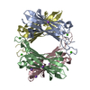
| ||||||||
|---|---|---|---|---|---|---|---|---|---|
| 1 | 
| ||||||||
| 2 | 
| ||||||||
| 3 | 
| ||||||||
| 4 | 
| ||||||||
| Unit cell |
|
-Components
| #1: Protein | Mass: 16058.631 Da / Num. of mol.: 4 Source method: isolated from a genetically manipulated source Source: (gene. exp.) Suipraeoptans intestinalis (bacteria) / Gene: FYJ34_05395 / Plasmid: pET28 / Production host: #2: Chemical | ChemComp-CA /   Mass: 40.078 Da / Num. of mol.: 4 / Source method: obtained synthetically / Formula: Ca / Feature type: SUBJECT OF INVESTIGATION Mass: 40.078 Da / Num. of mol.: 4 / Source method: obtained synthetically / Formula: Ca / Feature type: SUBJECT OF INVESTIGATION#3: Chemical | ChemComp-NA /   Mass: 22.990 Da / Num. of mol.: 6 / Source method: obtained synthetically / Formula: Na / Feature type: SUBJECT OF INVESTIGATION Mass: 22.990 Da / Num. of mol.: 6 / Source method: obtained synthetically / Formula: Na / Feature type: SUBJECT OF INVESTIGATION#4: Water | ChemComp-HOH / |  Mass: 18.015 Da / Num. of mol.: 235 / Source method: isolated from a natural source / Formula: H2O Mass: 18.015 Da / Num. of mol.: 235 / Source method: isolated from a natural source / Formula: H2OHas ligand of interest | Y | Has protein modification | Y | |
|---|
-Experimental details
-Experiment
| Experiment | Method: X-RAY DIFFRACTION / Number of used crystals: 1 |
|---|
- Sample preparation
| Crystal | Density Matthews: 2.06 Å3/Da / Density % sol: 40.25 % |
|---|---|
| Crystal grow | Temperature: 291 K / Method: vapor diffusion, hanging drop / Details: 0.1 M HEPES, 70% MPD |
-Data collection
| Diffraction | Mean temperature: 100 K / Serial crystal experiment: N |
|---|---|
| Diffraction source | Source: SYNCHROTRON / Site: Australian Synchrotron  / Beamline: MX2 / Wavelength: 0.95372 Å / Beamline: MX2 / Wavelength: 0.95372 Å |
| Detector | Type: DECTRIS EIGER X 16M / Detector: PIXEL / Date: Jul 7, 2022 |
| Radiation | Protocol: SINGLE WAVELENGTH / Monochromatic (M) / Laue (L): M / Scattering type: x-ray |
| Radiation wavelength | Wavelength: 0.95372 Å / Relative weight: 1 |
| Reflection | Resolution: 1.49→48.09 Å / Num. obs: 81829 / % possible obs: 96.6 % / Redundancy: 5.9 % / Biso Wilson estimate: 18.9 Å2 / CC1/2: 0.999 / Net I/σ(I): 10.5 |
| Reflection shell | Resolution: 1.49→1.52 Å / Redundancy: 5.4 % / Num. unique obs: 3553 / CC1/2: 0.35 / % possible all: 84.8 |
- Processing
| Software |
| |||||||||||||||||||||||||||||||||||||||||||||||||||||||||||||||||||||||||||||||||||||||||||||||||||||||||||||||||||||||||||||||||||||||||||||||||||||||||||||||||||||||||||||||||||||||||||||||||||||||||||||||||||||||||||||||||||||||
|---|---|---|---|---|---|---|---|---|---|---|---|---|---|---|---|---|---|---|---|---|---|---|---|---|---|---|---|---|---|---|---|---|---|---|---|---|---|---|---|---|---|---|---|---|---|---|---|---|---|---|---|---|---|---|---|---|---|---|---|---|---|---|---|---|---|---|---|---|---|---|---|---|---|---|---|---|---|---|---|---|---|---|---|---|---|---|---|---|---|---|---|---|---|---|---|---|---|---|---|---|---|---|---|---|---|---|---|---|---|---|---|---|---|---|---|---|---|---|---|---|---|---|---|---|---|---|---|---|---|---|---|---|---|---|---|---|---|---|---|---|---|---|---|---|---|---|---|---|---|---|---|---|---|---|---|---|---|---|---|---|---|---|---|---|---|---|---|---|---|---|---|---|---|---|---|---|---|---|---|---|---|---|---|---|---|---|---|---|---|---|---|---|---|---|---|---|---|---|---|---|---|---|---|---|---|---|---|---|---|---|---|---|---|---|---|---|---|---|---|---|---|---|---|---|---|---|---|---|---|---|---|---|
| Refinement | Method to determine structure: MOLECULAR REPLACEMENT / Resolution: 1.49→48.027 Å / Cor.coef. Fo:Fc: 0.97 / Cor.coef. Fo:Fc free: 0.955 / SU B: 3.621 / SU ML: 0.058 / Cross valid method: FREE R-VALUE / ESU R: 0.087 / ESU R Free: 0.078 Details: Hydrogens have been added in their riding positions
| |||||||||||||||||||||||||||||||||||||||||||||||||||||||||||||||||||||||||||||||||||||||||||||||||||||||||||||||||||||||||||||||||||||||||||||||||||||||||||||||||||||||||||||||||||||||||||||||||||||||||||||||||||||||||||||||||||||||
| Solvent computation | Ion probe radii: 0.8 Å / Shrinkage radii: 0.8 Å / VDW probe radii: 1.2 Å / Solvent model: MASK BULK SOLVENT | |||||||||||||||||||||||||||||||||||||||||||||||||||||||||||||||||||||||||||||||||||||||||||||||||||||||||||||||||||||||||||||||||||||||||||||||||||||||||||||||||||||||||||||||||||||||||||||||||||||||||||||||||||||||||||||||||||||||
| Displacement parameters | Biso mean: 21.476 Å2
| |||||||||||||||||||||||||||||||||||||||||||||||||||||||||||||||||||||||||||||||||||||||||||||||||||||||||||||||||||||||||||||||||||||||||||||||||||||||||||||||||||||||||||||||||||||||||||||||||||||||||||||||||||||||||||||||||||||||
| Refinement step | Cycle: LAST / Resolution: 1.49→48.027 Å
| |||||||||||||||||||||||||||||||||||||||||||||||||||||||||||||||||||||||||||||||||||||||||||||||||||||||||||||||||||||||||||||||||||||||||||||||||||||||||||||||||||||||||||||||||||||||||||||||||||||||||||||||||||||||||||||||||||||||
| Refine LS restraints |
| |||||||||||||||||||||||||||||||||||||||||||||||||||||||||||||||||||||||||||||||||||||||||||||||||||||||||||||||||||||||||||||||||||||||||||||||||||||||||||||||||||||||||||||||||||||||||||||||||||||||||||||||||||||||||||||||||||||||
| LS refinement shell | Refine-ID: X-RAY DIFFRACTION / Total num. of bins used: 20
|
