[English] 日本語
Yorodumi
- PDB-8fh0: Crystal structure of mutant Androgen Receptor ligand binding doma... -

+
Open data


ID or keywords:

Loading...

-
Basic information

Entry
Database: PDB / ID: 8fh0
TitleCrystal structure of mutant Androgen Receptor ligand binding domain H875Y/F877L/T878A with DHT
ComponentsAndrogen receptor
KeywordsGENE REGULATION / AR / Androgen receptor / DHT / steroid receptor
Function / homology
Function and homology information


male somatic sex determination / activation of prostate induction by androgen receptor signaling pathway / lateral sprouting involved in mammary gland duct morphogenesis / POU domain binding / negative regulation of integrin biosynthetic process / regulation of developmental growth / male genitalia morphogenesis / positive regulation of integrin biosynthetic process / tertiary branching involved in mammary gland duct morphogenesis / animal organ formation ...male somatic sex determination / activation of prostate induction by androgen receptor signaling pathway / lateral sprouting involved in mammary gland duct morphogenesis / POU domain binding / negative regulation of integrin biosynthetic process / regulation of developmental growth / male genitalia morphogenesis / positive regulation of integrin biosynthetic process / tertiary branching involved in mammary gland duct morphogenesis / animal organ formation / androgen binding / Leydig cell differentiation / regulation of systemic arterial blood pressure / epithelial cell morphogenesis / prostate gland growth / epithelial cell differentiation involved in prostate gland development / non-membrane-bounded organelle assembly / positive regulation of epithelial cell proliferation involved in prostate gland development / prostate gland epithelium morphogenesis / intracellular receptor signaling pathway / cellular response to testosterone stimulus / RNA polymerase II general transcription initiation factor binding / positive regulation of insulin-like growth factor receptor signaling pathway / positive regulation of transcription by RNA polymerase III / positive regulation of intracellular estrogen receptor signaling pathway / cellular response to steroid hormone stimulus / morphogenesis of an epithelial fold / seminiferous tubule development / androgen receptor signaling pathway / RUNX2 regulates osteoblast differentiation / single fertilization / mammary gland alveolus development / regulation of protein localization to plasma membrane / estrogen receptor signaling pathway / cellular response to estrogen stimulus / estrogen response element binding / nuclear receptor-mediated steroid hormone signaling pathway / positive regulation of phosphorylation / HSP90 chaperone cycle for steroid hormone receptors (SHR) in the presence of ligand / steroid binding / insulin-like growth factor receptor signaling pathway / molecular condensate scaffold activity / epithelial cell proliferation / G protein-coupled receptor activity / positive regulation of cell differentiation / negative regulation of extrinsic apoptotic signaling pathway / SUMOylation of intracellular receptors / Activated PKN1 stimulates transcription of AR (androgen receptor) regulated genes KLK2 and KLK3 / multicellular organism growth / beta-catenin binding / transcription coactivator binding / positive regulation of miRNA transcription / Nuclear Receptor transcription pathway / nuclear receptor activity / negative regulation of epithelial cell proliferation / male gonad development / MAPK cascade / cell-cell signaling / positive regulation of NF-kappaB transcription factor activity / ATPase binding / spermatogenesis / DNA-binding transcription activator activity, RNA polymerase II-specific / in utero embryonic development / RNA polymerase II-specific DNA-binding transcription factor binding / transcription by RNA polymerase II / positive regulation of MAPK cascade / molecular adaptor activity / transcription cis-regulatory region binding / DNA-binding transcription factor activity, RNA polymerase II-specific / Ub-specific processing proteases / nuclear speck / RNA polymerase II cis-regulatory region sequence-specific DNA binding / DNA-binding transcription factor activity / negative regulation of cell population proliferation / signaling receptor binding / chromatin binding / positive regulation of cell population proliferation / positive regulation of gene expression / chromatin / positive regulation of DNA-templated transcription / enzyme binding / negative regulation of transcription by RNA polymerase II / signal transduction / positive regulation of transcription by RNA polymerase II / protein-containing complex / zinc ion binding / nucleoplasm / nucleus / plasma membrane / cytoplasm / cytosol
Similarity search - Function
Androgen receptor / Androgen receptor / Nuclear hormones receptors DNA-binding region signature. / Zinc finger, nuclear hormone receptor-type / Zinc finger, C4 type (two domains) / Nuclear hormone receptors DNA-binding domain profile. / c4 zinc finger in nuclear hormone receptors / Nuclear hormone receptor, ligand-binding domain / Nuclear hormone receptor-like domain superfamily / Ligand-binding domain of nuclear hormone receptor ...Androgen receptor / Androgen receptor / Nuclear hormones receptors DNA-binding region signature. / Zinc finger, nuclear hormone receptor-type / Zinc finger, C4 type (two domains) / Nuclear hormone receptors DNA-binding domain profile. / c4 zinc finger in nuclear hormone receptors / Nuclear hormone receptor, ligand-binding domain / Nuclear hormone receptor-like domain superfamily / Ligand-binding domain of nuclear hormone receptor / Nuclear receptor (NR) ligand-binding (LBD) domain profile. / Ligand binding domain of hormone receptors / Zinc finger, NHR/GATA-type
Similarity search - Domain/homology
5-ALPHA-DIHYDROTESTOSTERONE / Androgen receptor
Similarity search - Component
Biological speciesHomo sapiens (human)
MethodX-RAY DIFFRACTION / SYNCHROTRON / MOLECULAR REPLACEMENT / Resolution: 1.59 Å
AuthorsDoamekpor, S.K. / Tong, L.
Funding support1items
OrganizationGrant numberCountry
Other private
CitationJournal: Acta Crystallogr.,Sect.F / Year: 2023
Title: A partially open conformation of an androgen receptor ligand-binding domain with drug-resistance mutations.
Authors: Doamekpor, S.K. / Peng, P. / Xu, R. / Ma, L. / Tong, Y. / Tong, L.
History
DepositionDec 13, 2022Deposition site: RCSB / Processing site: RCSB
Revision 1.0Apr 12, 2023Provider: repository / Type: Initial release
Revision 1.1May 22, 2024Group: Data collection / Category: chem_comp_atom / chem_comp_bond

-
Structure visualization

Structure viewerMolecule:
MolmilJmol/JSmol

Downloads & links

-
Assembly

Deposited unit
A: Androgen receptor
hetero molecules


Theoretical massNumber of molelcules
Total (without water)30,4423
Polymers30,0551
Non-polymers3872
Water1,946108
1


  • Idetical with deposited unit
  • defined by author&software
  • Evidence: gel filtration
TypeNameSymmetry operationNumber
identity operation1_555x,y,z1
MethodPISA
Unit cell
Length a, b, c (Å)56.522, 65.979, 73.136
Angle α, β, γ (deg.)90.00, 90.00, 90.00
Int Tables number19
Space group name H-MP212121

-
Components

#1: Protein Androgen receptor / Dihydrotestosterone receptor / Nuclear receptor subfamily 3 group C member 4


Mass: 30055.156 Da / Num. of mol.: 1 / Mutation: H875Y,F877L,T878A
Source method: isolated from a genetically manipulated source
Source: (gene. exp.) Homo sapiens (human) / Gene: AR, DHTR, NR3C4 / Production host: Escherichia coli (E. coli) / References: UniProt: P10275
#2: Chemical ChemComp-DHT / 5-ALPHA-DIHYDROTESTOSTERONE


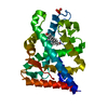
Mass: 290.440 Da / Num. of mol.: 1 / Source method: obtained synthetically / Formula: C19H30O2 / Feature type: SUBJECT OF INVESTIGATION / Comment: hormone*YM
#3: Chemical ChemComp-SO4 / SULFATE ION


Mass: 96.063 Da / Num. of mol.: 1 / Source method: obtained synthetically / Formula: SO4
#4: Water ChemComp-HOH / water


Mass: 18.015 Da / Num. of mol.: 108 / Source method: isolated from a natural source / Formula: H2O
Has ligand of interestY

-
Experimental details

-
Experiment

ExperimentMethod: X-RAY DIFFRACTION / Number of used crystals: 1

-
Sample preparation

CrystalDensity Matthews: 2.27 Å3/Da / Density % sol: 45.78 %
Crystal growTemperature: 293 K / Method: vapor diffusion, sitting drop
Details: 0.4-0.8 M sodium citrate, 0.1 M Hepes pH 7.5, 20% (v/v) ethylene glycol

-
Data collection

DiffractionMean temperature: 100 K / Serial crystal experiment: N
Diffraction sourceSource: SYNCHROTRON / Site: APS / Beamline: 24-ID-C / Wavelength: 0.9792 Å
DetectorType: DECTRIS EIGER X 16M / Detector: PIXEL / Date: Oct 7, 2021
RadiationProtocol: SINGLE WAVELENGTH / Monochromatic (M) / Laue (L): M / Scattering type: x-ray
Radiation wavelengthWavelength: 0.9792 Å / Relative weight: 1
ReflectionResolution: 1.59→200 Å / Num. obs: 70466 / % possible obs: 99.7 % / Redundancy: 3.86 % / CC1/2: 0.999 / Rmerge(I) obs: 0.035 / Rrim(I) all: 0.04 / Net I/σ(I): 16.48
Reflection shell
Resolution (Å)Rmerge(I) obsNum. unique obsCC1/2Rrim(I) allDiffraction-ID
1.59-1.690.62113180.820.7221
1.69-1.810.321107490.9430.3771
1.81-1.950.17899920.9850.2051
1.95-2.140.08692020.9950.0991
2.14-2.390.05182850.9970.061
2.39-2.760.03872890.9980.0441
2.76-3.380.03362100.9990.0381
3.38-4.770.02747770.9990.0321
4.77-2000.02626440.9990.031

-
Processing

Software
NameVersionClassification
PHENIXv1.2refinement
XDSdata scaling
XDSdata reduction
PHASERphasing
RefinementMethod to determine structure: MOLECULAR REPLACEMENT / Resolution: 1.59→48.99 Å / SU ML: 0.18 / Cross valid method: FREE R-VALUE / σ(F): 1.35 / Phase error: 25.44 / Stereochemistry target values: ML
RfactorNum. reflection% reflection
Rfree0.2082 2000 5.38 %
Rwork0.1957 --
obs0.1964 37154 99.81 %
Solvent computationShrinkage radii: 0.9 Å / VDW probe radii: 1.1 Å / Solvent model: FLAT BULK SOLVENT MODEL
Refinement stepCycle: LAST / Resolution: 1.59→48.99 Å
ProteinNucleic acidLigandSolventTotal
Num. atoms1967 0 26 108 2101
Refine LS restraints
Refine-IDTypeDev idealNumber
X-RAY DIFFRACTIONf_bond_d0.0062050
X-RAY DIFFRACTIONf_angle_d0.9312776
X-RAY DIFFRACTIONf_dihedral_angle_d15.204760
X-RAY DIFFRACTIONf_chiral_restr0.066306
X-RAY DIFFRACTIONf_plane_restr0.01346
LS refinement shell
Resolution (Å)Rfactor RfreeNum. reflection RfreeRfactor RworkNum. reflection RworkRefine-ID% reflection obs (%)
1.59-1.630.29311380.30782426X-RAY DIFFRACTION99
1.63-1.680.27181410.26872482X-RAY DIFFRACTION100
1.68-1.730.27321400.25252471X-RAY DIFFRACTION100
1.73-1.780.31321430.27092499X-RAY DIFFRACTION100
1.78-1.850.26331400.25812465X-RAY DIFFRACTION100
1.85-1.920.23421420.23022488X-RAY DIFFRACTION100
1.92-2.010.23121420.20312496X-RAY DIFFRACTION100
2.01-2.110.24041420.20242494X-RAY DIFFRACTION100
2.11-2.250.25631430.2012502X-RAY DIFFRACTION100
2.25-2.420.2071420.19712510X-RAY DIFFRACTION100
2.42-2.660.20211430.20492524X-RAY DIFFRACTION100
2.66-3.050.20261450.2132536X-RAY DIFFRACTION100
3.05-3.840.19991460.18772579X-RAY DIFFRACTION100
3.84-48.990.18241530.16772682X-RAY DIFFRACTION100

+
About Yorodumi

-
News

-
Feb 9, 2022. New format data for meta-information of EMDB entries

New format data for meta-information of EMDB entries

  • Version 3 of the EMDB header file is now the official format.
  • The previous official version 1.9 will be removed from the archive.

Related info.:EMDB header

External links:wwPDB to switch to version 3 of the EMDB data model

-
Aug 12, 2020. Covid-19 info

Covid-19 info

URL: https://pdbjlvh1.pdbj.org/emnavi/covid19.php

New page: Covid-19 featured information page in EM Navigator.

Related info.:Covid-19 info / Mar 5, 2020. Novel coronavirus structure data

+
Mar 5, 2020. Novel coronavirus structure data

Novel coronavirus structure data

Related info.:Yorodumi Speices / Aug 12, 2020. Covid-19 info

External links:COVID-19 featured content - PDBj / Molecule of the Month (242):Coronavirus Proteases

+
Jan 31, 2019. EMDB accession codes are about to change! (news from PDBe EMDB page)

EMDB accession codes are about to change! (news from PDBe EMDB page)

  • The allocation of 4 digits for EMDB accession codes will soon come to an end. Whilst these codes will remain in use, new EMDB accession codes will include an additional digit and will expand incrementally as the available range of codes is exhausted. The current 4-digit format prefixed with “EMD-” (i.e. EMD-XXXX) will advance to a 5-digit format (i.e. EMD-XXXXX), and so on. It is currently estimated that the 4-digit codes will be depleted around Spring 2019, at which point the 5-digit format will come into force.
  • The EM Navigator/Yorodumi systems omit the EMD- prefix.

Related info.:Q: What is EMD? / ID/Accession-code notation in Yorodumi/EM Navigator

External links:EMDB Accession Codes are Changing Soon! / Contact to PDBj

+
Jul 12, 2017. Major update of PDB

Major update of PDB

  • wwPDB released updated PDB data conforming to the new PDBx/mmCIF dictionary.
  • This is a major update changing the version number from 4 to 5, and with Remediation, in which all the entries are updated.
  • In this update, many items about electron microscopy experimental information are reorganized (e.g. em_software).
  • Now, EM Navigator and Yorodumi are based on the updated data.

External links:wwPDB Remediation / Enriched Model Files Conforming to OneDep Data Standards Now Available in the PDB FTP Archive

-
Yorodumi

Thousand views of thousand structures

  • Yorodumi is a browser for structure data from EMDB, PDB, SASBDB, etc.
  • This page is also the successor to EM Navigator detail page, and also detail information page/front-end page for Omokage search.
  • The word "yorodu" (or yorozu) is an old Japanese word meaning "ten thousand". "mi" (miru) is to see.

Related info.:EMDB / PDB / SASBDB / Comparison of 3 databanks / Yorodumi Search / Aug 31, 2016. New EM Navigator & Yorodumi / Yorodumi Papers / Jmol/JSmol / Function and homology information / Changes in new EM Navigator and Yorodumi

Read more