 ムービー
ムービー コントローラー


+ データを開く

- 基本情報
基本情報
| 登録情報 | データベース: PDB / ID: 8fg2 | ||||||||||||
|---|---|---|---|---|---|---|---|---|---|---|---|---|---|
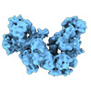
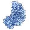
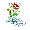
| タイトル | SARS-CoV-2 Nucleocapsid dimer structure determined from COVID-19 patients | ||||||||||||
 要素 要素 | Nucleoprotein | ||||||||||||
 キーワード キーワード | VIRAL PROTEIN / SARS-CoV-2 / N protein / COVID-19 / RNA binding protein | ||||||||||||
| 機能・相同性 |  機能・相同性情報 機能・相同性情報cytoplasmic capsid assembly / viral RNA genome packaging / response to host immune response / negative regulation of interferon-beta production / Maturation of nucleoprotein / intracellular non-membrane-bounded organelle / positive regulation of NLRP3 inflammasome complex assembly / MHC class I protein binding / CD28 dependent PI3K/Akt signaling / SARS-CoV-2 targets host intracellular signalling and regulatory pathways ...cytoplasmic capsid assembly / viral RNA genome packaging / response to host immune response / negative regulation of interferon-beta production / Maturation of nucleoprotein / intracellular non-membrane-bounded organelle / positive regulation of NLRP3 inflammasome complex assembly / MHC class I protein binding / CD28 dependent PI3K/Akt signaling / SARS-CoV-2 targets host intracellular signalling and regulatory pathways / protein sequestering activity / VEGFR2 mediated vascular permeability / molecular condensate scaffold activity / TAK1-dependent IKK and NF-kappa-B activation / NOD1/2 Signaling Pathway / DDX58/IFIH1-mediated induction of interferon-alpha/beta / MHC class I protein complex / Interleukin-1 signaling / RNA stem-loop binding / Interferon alpha/beta signaling / viral capsid / PIP3 activates AKT signaling / Transcription of SARS-CoV-2 sgRNAs / host cell endoplasmic reticulum-Golgi intermediate compartment / host cell Golgi apparatus / viral nucleocapsid / Translation of Structural Proteins / Virion Assembly and Release / host extracellular space / Induction of Cell-Cell Fusion / Attachment and Entry / host cell perinuclear region of cytoplasm / ribonucleoprotein complex / SARS-CoV-2 activates/modulates innate and adaptive immune responses / protein homodimerization activity / RNA binding / extracellular region / identical protein binding / cytoplasm 類似検索 - 分子機能 | ||||||||||||
| 生物種 |  Homo sapiens (ヒト) Homo sapiens (ヒト) | ||||||||||||
| 手法 | 電子顕微鏡法 / 単粒子再構成法 / クライオ電子顕微鏡法 / 解像度: 6 Å | ||||||||||||
 データ登録者 データ登録者 | Casasanta, M. / Jonaid, G.M. / Kaylor, L. / Luqiu, W. / DiCecco, L. / Solares, M. / Berry, S. / Kelly, D.F. | ||||||||||||
| 資金援助 |  米国, 3件 米国, 3件
| ||||||||||||
 引用 引用 | ジャーナル: Microsc Microanal / 年: 2023 タイトル: Structural Insights of the SARS-CoV-2 Nucleocapsid Protein: Implications for the Inner-workings of Rapid Antigen Tests. 著者: Michael A Casasanta / G M Jonaid / Liam Kaylor / William Y Luqiu / Liza-Anastasia DiCecco / Maria J Solares / Samantha Berry / William J Dearnaley / Deborah F Kelly /  要旨: The nucleocapsid (N) protein is an abundant component of SARS-CoV-2 and a key analyte for lateral-flow rapid antigen tests. Here, we present new structural insights for the SARS-CoV-2 N protein using ...The nucleocapsid (N) protein is an abundant component of SARS-CoV-2 and a key analyte for lateral-flow rapid antigen tests. Here, we present new structural insights for the SARS-CoV-2 N protein using cryo-electron microscopy (EM) and molecular modeling tools. Epitope mapping based on structural data supported host-immune interactions in the C-terminal portion of the protein, while other regions revealed protein-protein interaction sites. Complementary modeling results suggested that N protein structures from known variants of concern (VOC) are nearly 100% conserved at specific antibody-binding sites. Collectively, these results suggest that rapid tests that target the nucleocapsid C-terminal domain should have similar accuracy across all VOCs. In addition, our combined structural modeling workflow may guide the design of immune therapies to counter viral processes as we plan for future variants and pandemics. #1: ジャーナル: Microsc Microanal / 年: 2022タイトル: Structural insights of the SARS-CoV-2 Nucleocapsid protein: Implications for the inner-workings of rapid antigen tests 著者: Casasanta, M. / Jonaid, G.M. / Kaylor, L. / Luqiu, W. / DiCecco, L. / Solares, M. / Berry, S. / Kelly, D.F. #2: ジャーナル: Nanoscale / 年: 2021タイトル: Microchip-based structure determination of low-molecular weight proteins using cryo-electron microscopy. 著者: Michael A Casasanta / G M Jonaid / Liam Kaylor / William Y Luqiu / Maria J Solares / Mariah L Schroen / William J Dearnaley / Jarad Wilson / Madeline J Dukes / Deborah F Kelly / 要旨: Interest in cryo-Electron Microscopy (EM) imaging has skyrocketed in recent years due to its pristine views of macromolecules and materials. As advances in instrumentation and computing algorithms ...Interest in cryo-Electron Microscopy (EM) imaging has skyrocketed in recent years due to its pristine views of macromolecules and materials. As advances in instrumentation and computing algorithms spurred this progress, there is renewed focus to address specimen-related challenges. Here we contribute a microchip-based toolkit to perform complementary structural and biochemical analysis on low-molecular weight proteins. As a model system, we used the SARS-CoV-2 nucleocapsid (N) protein (48 kDa) due to its stability and important role in therapeutic development. Cryo-EM structures of the N protein monomer revealed a flexible N-terminal "top hat" motif and a helical-rich C-terminal domain. To complement our structural findings, we engineered microchip-based immunoprecipitation assays that led to the discovery of the first antibody binding site on the N protein. The data also facilitated molecular modeling of a variety of pandemic and common cold-related coronavirus proteins. Such insights may guide future pandemic-preparedness protocols through immuno-engineering strategies to mitigate viral outbreaks. | ||||||||||||
| 履歴 |
|
- 構造の表示
構造の表示
| 構造ビューア | 分子: MolmilJmol/JSmol |
|---|
- ダウンロードとリンク
-ダウンロード
| PDBx/mmCIF形式 | 8fg2.cif.gz | 146.2 KB | 表示 | PDBx/mmCIF形式 |
|---|---|---|---|---|
| PDB形式 | pdb8fg2.ent.gz | 115.5 KB | 表示 | PDB形式 |
| PDBx/mmJSON形式 | 8fg2.json.gz | ツリー表示 | PDBx/mmJSON形式 | |
| その他 |  その他のダウンロード その他のダウンロード |
-検証レポート
| 文書・要旨 | 8fg2_validation.pdf.gz | 972.8 KB | 表示 | wwPDB検証レポート |
|---|---|---|---|---|
| 文書・詳細版 | 8fg2_full_validation.pdf.gz | 975.3 KB | 表示 | |
| XML形式データ | 8fg2_validation.xml.gz | 29 KB | 表示 | |
| CIF形式データ | 8fg2_validation.cif.gz | 43.9 KB | 表示 | |
| アーカイブディレクトリ | https://data.pdbj.org/pub/pdb/validation_reports/fg/8fg2ftp://data.pdbj.org/pub/pdb/validation_reports/fg/8fg2 | HTTPS FTP |
-関連構造データ
-リンク
 PDBj
PDBj


- 集合体
集合体
| 登録構造単位 | 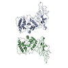
|
|---|---|
| 1 |
|
-要素
| #1: タンパク質 | 分子量: 45689.645 Da / 分子数: 2 / 由来タイプ: 天然 / 由来: (天然) Homo sapiens (ヒト) / 参照: UniProt: P0DTC9 |
|---|
-実験情報
-実験
| 実験 | 手法: 電子顕微鏡法 |
|---|---|
| EM実験 | 試料の集合状態: PARTICLE / 3次元再構成法: 単粒子再構成法 |
- 試料調製
試料調製
| 構成要素 | 名称: Nucleocapsid dimer is comprised of A chain and B chain タイプ: COMPLEX 詳細: For each monomer that comprises the dimer structure, residues 1-49 were fit into the map separately from residues 50 - 419. Entity ID: all / 由来: NATURAL |
|---|---|
| 分子量 | 実験値: NO |
| 由来(天然) | 生物種:   Severe acute respiratory syndrome coronavirus 2 (ウイルス) Severe acute respiratory syndrome coronavirus 2 (ウイルス) |
| 由来(組換発現) | 生物種:  |
| 緩衝液 | pH: 7.5 詳細: 20 mM Tris (pH 7.5), 150 mM NaCl, 10 mM MgCl2, 10 mM CaCl2 |
| 試料 | 濃度: 0.1 mg/ml / 包埋: NO / シャドウイング: NO / 染色: NO / 凍結: YES 詳細: Sample was enriched using Ni-NTA coated silicon nitride microchips |
| 試料支持 | 詳細: Samples were incubated with Ni-NTA coated microchips for 1 minute prior to plunge freezing into liquid ethane. グリッドの材料: SILICON NITRIDE / グリッドのタイプ: Homemade |
| 急速凍結 | 装置: FEI VITROBOT MARK III / 凍結剤: ETHANE / 湿度: 100 % / 凍結前の試料温度: 298 K 詳細: A Mark III Vitrobot was used to plunge samples into liquid ethane, operating at room temperature and 100% humidity with 3 - 4 seconds blot time |
- 電子顕微鏡撮影
| 実験機器 |  モデル: Titan Krios / 画像提供: FEI Company |
|---|---|
| 顕微鏡 | モデル: TFS KRIOS |
| 電子銃 | 電子線源:  FIELD EMISSION GUN / 加速電圧: 300 kV / 照射モード: FLOOD BEAM FIELD EMISSION GUN / 加速電圧: 300 kV / 照射モード: FLOOD BEAM |
| 電子レンズ | モード: BRIGHT FIELD / 倍率(公称値): 59000 X / 最大 デフォーカス(公称値): 3000 nm / 最小 デフォーカス(公称値): 1000 nm / アライメント法: BASIC |
| 試料ホルダ | 凍結剤: NITROGEN 試料ホルダーモデル: FEI TITAN KRIOS AUTOGRID HOLDER |
| 撮影 | 平均露光時間: 1 sec. / 電子線照射量: 50 e/Å2 / 検出モード: INTEGRATING フィルム・検出器のモデル: FEI FALCON III (4k x 4k) 撮影したグリッド数: 4 / 実像数: 300 |
| 画像スキャン | サンプリングサイズ: 14 µm / 動画フレーム数/画像: 30 |
- 解析
解析
| ソフトウェア | 名称: PHENIX / バージョン: 1.17.1_3660: / 分類: 精密化 | ||||||||||||||||||||||||||||||
|---|---|---|---|---|---|---|---|---|---|---|---|---|---|---|---|---|---|---|---|---|---|---|---|---|---|---|---|---|---|---|---|
| EMソフトウェア |
| ||||||||||||||||||||||||||||||
| CTF補正 | タイプ: PHASE FLIPPING ONLY | ||||||||||||||||||||||||||||||
| 粒子像の選択 | 選択した粒子像数: 10000 | ||||||||||||||||||||||||||||||
| 対称性 | 点対称性: C1 (非対称) | ||||||||||||||||||||||||||||||
| 3次元再構成 | 解像度: 6 Å / 解像度の算出法: FSC 0.143 CUT-OFF / 粒子像の数: 10000 / クラス平均像の数: 1 / 対称性のタイプ: POINT | ||||||||||||||||||||||||||||||
| 原子モデル構築 | B value: 100 / プロトコル: RIGID BODY FIT / 空間: REAL | ||||||||||||||||||||||||||||||
| 拘束条件 |
|
