 Movie
Movie Controller

[English] 日本語
 Yorodumi
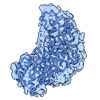
- PDB-8fg2: SARS-CoV-2 Nucleocapsid dimer structure determined from COVID-19 ... -
+ Open data

- Basic information
| Entry | Database: PDB / ID: 8fg2 | ||||||||||||
|---|---|---|---|---|---|---|---|---|---|---|---|---|---|
| Title | SARS-CoV-2 Nucleocapsid dimer structure determined from COVID-19 patients | ||||||||||||
 Components Components | Nucleoprotein | ||||||||||||
 Keywords Keywords | VIRAL PROTEIN / SARS-CoV-2 / N protein / COVID-19 / RNA binding protein | ||||||||||||
| Function / homology |  Function and homology information Function and homology informationcytoplasmic capsid assembly / viral RNA genome packaging / response to host immune response / negative regulation of interferon-beta production / Maturation of nucleoprotein / intracellular non-membrane-bounded organelle / positive regulation of NLRP3 inflammasome complex assembly / MHC class I protein binding / CD28 dependent PI3K/Akt signaling / SARS-CoV-2 targets host intracellular signalling and regulatory pathways ...cytoplasmic capsid assembly / viral RNA genome packaging / response to host immune response / negative regulation of interferon-beta production / Maturation of nucleoprotein / intracellular non-membrane-bounded organelle / positive regulation of NLRP3 inflammasome complex assembly / MHC class I protein binding / CD28 dependent PI3K/Akt signaling / SARS-CoV-2 targets host intracellular signalling and regulatory pathways / protein sequestering activity / VEGFR2 mediated vascular permeability / molecular condensate scaffold activity / TAK1-dependent IKK and NF-kappa-B activation / NOD1/2 Signaling Pathway / DDX58/IFIH1-mediated induction of interferon-alpha/beta / MHC class I protein complex / Interleukin-1 signaling / RNA stem-loop binding / Interferon alpha/beta signaling / viral capsid / PIP3 activates AKT signaling / Transcription of SARS-CoV-2 sgRNAs / host cell endoplasmic reticulum-Golgi intermediate compartment / host cell Golgi apparatus / viral nucleocapsid / Translation of Structural Proteins / Virion Assembly and Release / host extracellular space / Induction of Cell-Cell Fusion / Attachment and Entry / host cell perinuclear region of cytoplasm / ribonucleoprotein complex / SARS-CoV-2 activates/modulates innate and adaptive immune responses / protein homodimerization activity / RNA binding / extracellular region / identical protein binding / cytoplasm Similarity search - Function | ||||||||||||
| Biological species |  Homo sapiens (human) Homo sapiens (human) | ||||||||||||
| Method | ELECTRON MICROSCOPY / single particle reconstruction / cryo EM / Resolution: 6 Å | ||||||||||||
 Authors Authors | Casasanta, M. / Jonaid, G.M. / Kaylor, L. / Luqiu, W. / DiCecco, L. / Solares, M. / Berry, S. / Kelly, D.F. | ||||||||||||
| Funding support |  United States, 3items United States, 3items
| ||||||||||||
 Citation Citation | Journal: Microsc Microanal / Year: 2023 Title: Structural Insights of the SARS-CoV-2 Nucleocapsid Protein: Implications for the Inner-workings of Rapid Antigen Tests. Authors: Michael A Casasanta / G M Jonaid / Liam Kaylor / William Y Luqiu / Liza-Anastasia DiCecco / Maria J Solares / Samantha Berry / William J Dearnaley / Deborah F Kelly /  Abstract: The nucleocapsid (N) protein is an abundant component of SARS-CoV-2 and a key analyte for lateral-flow rapid antigen tests. Here, we present new structural insights for the SARS-CoV-2 N protein using ...The nucleocapsid (N) protein is an abundant component of SARS-CoV-2 and a key analyte for lateral-flow rapid antigen tests. Here, we present new structural insights for the SARS-CoV-2 N protein using cryo-electron microscopy (EM) and molecular modeling tools. Epitope mapping based on structural data supported host-immune interactions in the C-terminal portion of the protein, while other regions revealed protein-protein interaction sites. Complementary modeling results suggested that N protein structures from known variants of concern (VOC) are nearly 100% conserved at specific antibody-binding sites. Collectively, these results suggest that rapid tests that target the nucleocapsid C-terminal domain should have similar accuracy across all VOCs. In addition, our combined structural modeling workflow may guide the design of immune therapies to counter viral processes as we plan for future variants and pandemics. #1: Journal: Microsc Microanal / Year: 2022Title: Structural insights of the SARS-CoV-2 Nucleocapsid protein: Implications for the inner-workings of rapid antigen tests Authors: Casasanta, M. / Jonaid, G.M. / Kaylor, L. / Luqiu, W. / DiCecco, L. / Solares, M. / Berry, S. / Kelly, D.F. #2: Journal: Nanoscale / Year: 2021Title: Microchip-based structure determination of low-molecular weight proteins using cryo-electron microscopy. Authors: Michael A Casasanta / G M Jonaid / Liam Kaylor / William Y Luqiu / Maria J Solares / Mariah L Schroen / William J Dearnaley / Jarad Wilson / Madeline J Dukes / Deborah F Kelly / Abstract: Interest in cryo-Electron Microscopy (EM) imaging has skyrocketed in recent years due to its pristine views of macromolecules and materials. As advances in instrumentation and computing algorithms ...Interest in cryo-Electron Microscopy (EM) imaging has skyrocketed in recent years due to its pristine views of macromolecules and materials. As advances in instrumentation and computing algorithms spurred this progress, there is renewed focus to address specimen-related challenges. Here we contribute a microchip-based toolkit to perform complementary structural and biochemical analysis on low-molecular weight proteins. As a model system, we used the SARS-CoV-2 nucleocapsid (N) protein (48 kDa) due to its stability and important role in therapeutic development. Cryo-EM structures of the N protein monomer revealed a flexible N-terminal "top hat" motif and a helical-rich C-terminal domain. To complement our structural findings, we engineered microchip-based immunoprecipitation assays that led to the discovery of the first antibody binding site on the N protein. The data also facilitated molecular modeling of a variety of pandemic and common cold-related coronavirus proteins. Such insights may guide future pandemic-preparedness protocols through immuno-engineering strategies to mitigate viral outbreaks. | ||||||||||||
| History |
|
- Structure visualization
| Structure viewer | Molecule: MolmilJmol/JSmol |
|---|
- Downloads & links
-Download
| PDBx/mmCIF format | 8fg2.cif.gz | 146.2 KB | Display | PDBx/mmCIF format |
|---|---|---|---|---|
| PDB format | pdb8fg2.ent.gz | 115.5 KB | Display | PDB format |
| PDBx/mmJSON format | 8fg2.json.gz | Tree view | PDBx/mmJSON format | |
| Others |  Other downloads Other downloads |
-Validation report
| Summary document | 8fg2_validation.pdf.gz | 972.8 KB | Display | wwPDB validaton report |
|---|---|---|---|---|
| Full document | 8fg2_full_validation.pdf.gz | 975.3 KB | Display | |
| Data in XML | 8fg2_validation.xml.gz | 29 KB | Display | |
| Data in CIF | 8fg2_validation.cif.gz | 43.9 KB | Display | |
| Arichive directory | https://data.pdbj.org/pub/pdb/validation_reports/fg/8fg2ftp://data.pdbj.org/pub/pdb/validation_reports/fg/8fg2 | HTTPS FTP |
-Related structure data
| Related structure data | 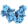 29072MC 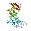 8fd5C M: map data used to model this data C: citing same article ( |
|---|---|
| Similar structure data |
-Links
 PDBj
PDBj


- Assembly
| Deposited unit | 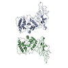
|
|---|---|
| 1 |
|
-Components
| #1: Protein | Mass: 45689.645 Da / Num. of mol.: 2 / Source method: isolated from a natural source / Source: (natural) Homo sapiens (human) / References: UniProt: P0DTC9 |
|---|
-Experimental details
-Experiment
| Experiment | Method: ELECTRON MICROSCOPY |
|---|---|
| EM experiment | Aggregation state: PARTICLE / 3D reconstruction method: single particle reconstruction |
- Sample preparation
| Component | Name: Nucleocapsid dimer is comprised of A chain and B chain Type: COMPLEX Details: For each monomer that comprises the dimer structure, residues 1-49 were fit into the map separately from residues 50 - 419. Entity ID: all / Source: NATURAL |
|---|---|
| Molecular weight | Experimental value: NO |
| Source (natural) | Organism:   Severe acute respiratory syndrome coronavirus 2 Severe acute respiratory syndrome coronavirus 2 |
| Source (recombinant) | Organism:  |
| Buffer solution | pH: 7.5 Details: 20 mM Tris (pH 7.5), 150 mM NaCl, 10 mM MgCl2, 10 mM CaCl2 |
| Specimen | Conc.: 0.1 mg/ml / Embedding applied: NO / Shadowing applied: NO / Staining applied: NO / Vitrification applied: YES Details: Sample was enriched using Ni-NTA coated silicon nitride microchips |
| Specimen support | Details: Samples were incubated with Ni-NTA coated microchips for 1 minute prior to plunge freezing into liquid ethane. Grid material: SILICON NITRIDE / Grid type: Homemade |
| Vitrification | Instrument: FEI VITROBOT MARK III / Cryogen name: ETHANE / Humidity: 100 % / Chamber temperature: 298 K Details: A Mark III Vitrobot was used to plunge samples into liquid ethane, operating at room temperature and 100% humidity with 3 - 4 seconds blot time |
- Electron microscopy imaging
| Experimental equipment |  Model: Titan Krios / Image courtesy: FEI Company |
|---|---|
| Microscopy | Model: TFS KRIOS |
| Electron gun | Electron source:  FIELD EMISSION GUN / Accelerating voltage: 300 kV / Illumination mode: FLOOD BEAM FIELD EMISSION GUN / Accelerating voltage: 300 kV / Illumination mode: FLOOD BEAM |
| Electron lens | Mode: BRIGHT FIELD / Nominal magnification: 59000 X / Nominal defocus max: 3000 nm / Nominal defocus min: 1000 nm / Alignment procedure: BASIC |
| Specimen holder | Cryogen: NITROGEN / Specimen holder model: FEI TITAN KRIOS AUTOGRID HOLDER |
| Image recording | Average exposure time: 1 sec. / Electron dose: 50 e/Å2 / Detector mode: INTEGRATING / Film or detector model: FEI FALCON III (4k x 4k) / Num. of grids imaged: 4 / Num. of real images: 300 |
| Image scans | Sampling size: 14 µm / Movie frames/image: 30 |
- Processing
| Software | Name: PHENIX / Version: 1.17.1_3660: / Classification: refinement | ||||||||||||||||||||||||||||||
|---|---|---|---|---|---|---|---|---|---|---|---|---|---|---|---|---|---|---|---|---|---|---|---|---|---|---|---|---|---|---|---|
| EM software |
| ||||||||||||||||||||||||||||||
| CTF correction | Type: PHASE FLIPPING ONLY | ||||||||||||||||||||||||||||||
| Particle selection | Num. of particles selected: 10000 | ||||||||||||||||||||||||||||||
| Symmetry | Point symmetry: C1 (asymmetric) | ||||||||||||||||||||||||||||||
| 3D reconstruction | Resolution: 6 Å / Resolution method: FSC 0.143 CUT-OFF / Num. of particles: 10000 / Num. of class averages: 1 / Symmetry type: POINT | ||||||||||||||||||||||||||||||
| Atomic model building | B value: 100 / Protocol: RIGID BODY FIT / Space: REAL | ||||||||||||||||||||||||||||||
| Refine LS restraints |
|
