 Movie
Movie Controller

[English] 日本語
 Yorodumi
- PDB-8fdb: CRYSTAL STRUCTURE OF NAGB-II PHOSPHOSUGAR ISOMERASE FROM Shewanel... -
+ Open data

- Basic information
| Entry | Database: PDB / ID: 8fdb | |||||||||
|---|---|---|---|---|---|---|---|---|---|---|
| Title | CRYSTAL STRUCTURE OF NAGB-II PHOSPHOSUGAR ISOMERASE FROM Shewanella denitrificans OS217 IN COMPLEX WITH GLUCITOLAMINE-6-PHOSPHATE AT 3.06 A RESOLUTION. | |||||||||
 Components Components | (Glutamine-fructose-6-phosphate transaminase ...) x 2 | |||||||||
 Keywords Keywords | ISOMERASE / Deaminase Isomerization-deamination Amino-sugar metabolism Positive cooperativity and allosteric activation Sugar-isomerase domain | |||||||||
| Function / homology |  Function and homology information Function and homology informationglutamine-fructose-6-phosphate transaminase (isomerizing) / L-glutamine:D-fructose-6-phosphate transaminase (isomerizing) activity / carbohydrate derivative metabolic process / carbohydrate derivative binding Similarity search - Function | |||||||||
| Biological species |  Shewanella denitrificans OS217 (bacteria) Shewanella denitrificans OS217 (bacteria) | |||||||||
| Method |  X-RAY DIFFRACTION / MOLECULAR REPLACEMENT / Resolution: 3.06 Å X-RAY DIFFRACTION / MOLECULAR REPLACEMENT / Resolution: 3.06 Å | |||||||||
 Authors Authors | Rodriguez-Romero, A. / Rodriguez-Hernandez, A. / Marcos-Viquez, J. / Bustos-Jaimes, I. | |||||||||
| Funding support |  Mexico, 2items Mexico, 2items
| |||||||||
 Citation Citation | Journal: Protein Sci. / Year: 2023 Title: Substrate binding in the allosteric site mimics homotropic cooperativity in the SIS-fold glucosamine-6-phosphate deaminases. Authors: Marcos-Viquez, J. / Rodriguez-Hernandez, A. / Alvarez-Anorve, L.I. / Medina-Garcia, A. / Plumbridge, J. / Calcagno, M.L. / Rodriguez-Romero, A. / Bustos-Jaimes, I. | |||||||||
| History |
|
- Structure visualization
| Structure viewer | Molecule: MolmilJmol/JSmol |
|---|
- Downloads & links
-Download
| PDBx/mmCIF format | 8fdb.cif.gz | 133 KB | Display | PDBx/mmCIF format |
|---|---|---|---|---|
| PDB format | pdb8fdb.ent.gz | 105.2 KB | Display | PDB format |
| PDBx/mmJSON format | 8fdb.json.gz | Tree view | PDBx/mmJSON format | |
| Others |  Other downloads Other downloads |
-Validation report
| Arichive directory | https://data.pdbj.org/pub/pdb/validation_reports/fd/8fdbftp://data.pdbj.org/pub/pdb/validation_reports/fd/8fdb | HTTPS FTP |
|---|
-Related structure data


-Links
 PDBj
PDBj- Assembly
| Deposited unit | 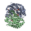
| ||||||||||
|---|---|---|---|---|---|---|---|---|---|---|---|
| 1 |
| ||||||||||
| Unit cell |
|
-Components
-Glutamine-fructose-6-phosphate transaminase ... , 2 types, 2 molecules AB
| #1: Protein | Mass: 35476.961 Da / Num. of mol.: 1 Source method: isolated from a genetically manipulated source Source: (gene. exp.) Shewanella denitrificans OS217 (bacteria)Strain: OS217 / ATCC BAA-1090 / DSM 15013 / Gene: Sden_2705 / Plasmid: PET24B / Production host: |
|---|---|
| #2: Protein | Mass: 35519.961 Da / Num. of mol.: 1 Source method: isolated from a genetically manipulated source Source: (gene. exp.) Shewanella denitrificans OS217 (bacteria)Gene: Sden_2705 / Plasmid: PET24B / Production host: |
-Non-polymers , 4 types, 24 molecules 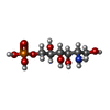


| #3: Chemical | ChemComp-AGP /  Mass: 261.167 Da / Num. of mol.: 4 / Source method: obtained synthetically / Formula: C6H16NO8P / Feature type: SUBJECT OF INVESTIGATION Mass: 261.167 Da / Num. of mol.: 4 / Source method: obtained synthetically / Formula: C6H16NO8P / Feature type: SUBJECT OF INVESTIGATION#4: Chemical |  Mass: 24.305 Da / Num. of mol.: 2 / Source method: obtained synthetically / Formula: Mg Mass: 24.305 Da / Num. of mol.: 2 / Source method: obtained synthetically / Formula: Mg#5: Chemical |  Mass: 92.094 Da / Num. of mol.: 2 / Source method: obtained synthetically / Formula: C3H8O3 Mass: 92.094 Da / Num. of mol.: 2 / Source method: obtained synthetically / Formula: C3H8O3#6: Water | ChemComp-HOH / | Mass: 18.015 Da / Num. of mol.: 16 / Source method: isolated from a natural source / Formula: H2O |
|---|
-Details
| Has ligand of interest | Y |
|---|
-Experimental details
-Experiment
| Experiment | Method: X-RAY DIFFRACTION / Number of used crystals: 1 |
|---|
- Sample preparation
| Crystal | Density Matthews: 2.45 Å3/Da / Density % sol: 49.86 % / Description: Bypyramidal plates |
|---|---|
| Crystal grow | Temperature: 291 K / Method: vapor diffusion, sitting drop / pH: 8.5 Details: 50mM Tris-HCl, pH 8.5, with 1mM 2-deoxi-2-amino glucitol 6-phosphate (GlcNol6P) |
-Data collection
| Diffraction | Mean temperature: 100 K / Serial crystal experiment: N |
|---|---|
| Diffraction source | Source: ROTATING ANODE / Type: RIGAKU MICROMAX-007 HF / Wavelength: 1.5419 Å |
| Detector | Type: DECTRIS PILATUS 200K / Detector: PIXEL / Date: Sep 17, 2018 / Details: Mirrors |
| Radiation | Protocol: SINGLE WAVELENGTH / Monochromatic (M) / Laue (L): M / Scattering type: x-ray |
| Radiation wavelength | Wavelength: 1.5419 Å / Relative weight: 1 |
| Reflection | Resolution: 3→49.48 Å / Num. obs: 14320 / % possible obs: 99.7 % / Redundancy: 5 % / Biso Wilson estimate: 45.49 Å2 / CC1/2: 0.794 / Rmerge(I) obs: 0.165 / Net I/σ(I): 9.8 |
| Reflection shell | Resolution: 3.06→3.27 Å / Redundancy: 3.2 % / Rmerge(I) obs: 0.389 / Mean I/σ(I) obs: 3 / Num. unique obs: 2383 / CC1/2: 0.89 / Rpim(I) all: 0.25 / Χ2: 0.82 / % possible all: 99.7 |
- Processing
| Software |
| |||||||||||||||||||||||||||||||||||||||||||||||||||||||||||||||
|---|---|---|---|---|---|---|---|---|---|---|---|---|---|---|---|---|---|---|---|---|---|---|---|---|---|---|---|---|---|---|---|---|---|---|---|---|---|---|---|---|---|---|---|---|---|---|---|---|---|---|---|---|---|---|---|---|---|---|---|---|---|---|---|---|
| Refinement | Method to determine structure: MOLECULAR REPLACEMENT Starting model: BALBES Resolution: 3.06→34.95 Å / SU ML: 0.35 / Cross valid method: THROUGHOUT / σ(F): 1.34 / Phase error: 24.16 / Stereochemistry target values: ML
| |||||||||||||||||||||||||||||||||||||||||||||||||||||||||||||||
| Solvent computation | Shrinkage radii: 0.9 Å / VDW probe radii: 1.1 Å / Solvent model: FLAT BULK SOLVENT MODEL | |||||||||||||||||||||||||||||||||||||||||||||||||||||||||||||||
| Refinement step | Cycle: LAST / Resolution: 3.06→34.95 Å
| |||||||||||||||||||||||||||||||||||||||||||||||||||||||||||||||
| Refine LS restraints |
| |||||||||||||||||||||||||||||||||||||||||||||||||||||||||||||||
| LS refinement shell |
|
