 Movie
Movie Controller

[English] 日本語
 Yorodumi
- PDB-8fbu: Crystal structure of Cryptosporidium parvum N-myristoyltransferas... -
+ Open data

- Basic information
| Entry | Database: PDB / ID: 8fbu | |||||||||
|---|---|---|---|---|---|---|---|---|---|---|
| Title | Crystal structure of Cryptosporidium parvum N-myristoyltransferase with bound myristoyl-CoA and Compound-2 | |||||||||
 Components Components | Glycylpeptide N-tetradecanoyltransferase | |||||||||
 Keywords Keywords | TRANSFERASE/INHIBITOR / NMT / inhibitor / Myristoyl-CoA / MyrCoA / Structural Genomics / Seattle Structural Genomics Center for Infectious Disease / SSGCID / TRANSFERASE / TRANSFERASE-INHIBITOR complex | |||||||||
| Function / homology |  Function and homology information Function and homology informationglycylpeptide N-tetradecanoyltransferase / glycylpeptide N-tetradecanoyltransferase activity / cytoplasm Similarity search - Function | |||||||||
| Biological species |  Cryptosporidium parvum Iowa II (eukaryote) Cryptosporidium parvum Iowa II (eukaryote) | |||||||||
| Method |  X-RAY DIFFRACTION / SYNCHROTRON / MOLECULAR REPLACEMENT / Resolution: 2 Å X-RAY DIFFRACTION / SYNCHROTRON / MOLECULAR REPLACEMENT / Resolution: 2 Å | |||||||||
 Authors Authors | Staker, B.L. / Fenwick, M.K. / Seattle Structural Genomics Center for Infectious Disease (SSGCID) | |||||||||
| Funding support |  United States, 2items United States, 2items
| |||||||||
 Citation Citation | Journal: Acs Infect Dis. / Year: 2023 Title: Identification of and Structural Insights into Hit Compounds Targeting N -Myristoyltransferase for Cryptosporidium Drug Development. Authors: Fenwick, M.K. / Reers, A.R. / Liu, Y. / Zigweid, R. / Sankaran, B. / Shin, J. / Hulverson, M.A. / Hammerson, B. / Fernandez Alvaro, E. / Myler, P.J. / Kaushansky, A. / Van Voorhis, W.C. / Fan, E. / Staker, B.L. | |||||||||
| History |
|
- Structure visualization
| Structure viewer | Molecule: MolmilJmol/JSmol |
|---|
- Downloads & links
-Download
| PDBx/mmCIF format | 8fbu.cif.gz | 554.8 KB | Display | PDBx/mmCIF format |
|---|---|---|---|---|
| PDB format | pdb8fbu.ent.gz | Display | PDB format | |
| PDBx/mmJSON format | 8fbu.json.gz | Tree view | PDBx/mmJSON format | |
| Others |  Other downloads Other downloads |
-Validation report
| Arichive directory | https://data.pdbj.org/pub/pdb/validation_reports/fb/8fbuftp://data.pdbj.org/pub/pdb/validation_reports/fb/8fbu | HTTPS FTP |
|---|
-Related structure data


-Links
 PDBj
PDBj

- Assembly
| Deposited unit | 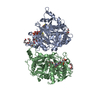
| ||||||||
|---|---|---|---|---|---|---|---|---|---|
| 1 | 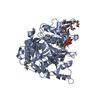
| ||||||||
| 2 | 
| ||||||||
| Unit cell |
|
-Components
-Protein , 1 types, 2 molecules AB
| #1: Protein | Mass: 50717.109 Da / Num. of mol.: 2 Source method: isolated from a genetically manipulated source Source: (gene. exp.) Cryptosporidium parvum Iowa II (eukaryote)Gene: cgd3_320 Production host:  References: UniProt: Q5CV46 |
|---|
-Non-polymers , 8 types, 588 molecules 
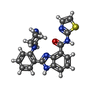


| #2: Chemical |  Mass: 977.890 Da / Num. of mol.: 2 / Source method: obtained synthetically / Formula: C35H62N7O17P3S / Feature type: SUBJECT OF INVESTIGATION Mass: 977.890 Da / Num. of mol.: 2 / Source method: obtained synthetically / Formula: C35H62N7O17P3S / Feature type: SUBJECT OF INVESTIGATION#3: Chemical | ChemComp-XOL / ( |  Mass: 404.488 Da / Num. of mol.: 1 / Source method: obtained synthetically / Formula: C21H20N6OS / Feature type: SUBJECT OF INVESTIGATION Mass: 404.488 Da / Num. of mol.: 1 / Source method: obtained synthetically / Formula: C21H20N6OS / Feature type: SUBJECT OF INVESTIGATION#4: Chemical |  Mass: 194.226 Da / Num. of mol.: 2 / Source method: obtained synthetically / Formula: C8H18O5 / Comment: precipitant*YM Mass: 194.226 Da / Num. of mol.: 2 / Source method: obtained synthetically / Formula: C8H18O5 / Comment: precipitant*YM#5: Chemical |  Mass: 238.278 Da / Num. of mol.: 2 / Source method: obtained synthetically / Formula: C10H22O6 / Comment: precipitant*YM Mass: 238.278 Da / Num. of mol.: 2 / Source method: obtained synthetically / Formula: C10H22O6 / Comment: precipitant*YM#6: Chemical | ChemComp-PGE / |  Mass: 150.173 Da / Num. of mol.: 1 / Source method: obtained synthetically / Formula: C6H14O4 Mass: 150.173 Da / Num. of mol.: 1 / Source method: obtained synthetically / Formula: C6H14O4#7: Chemical |  Mass: 62.068 Da / Num. of mol.: 2 / Source method: obtained synthetically / Formula: C2H6O2 Mass: 62.068 Da / Num. of mol.: 2 / Source method: obtained synthetically / Formula: C2H6O2#8: Chemical | ChemComp-CL / |  Mass: 35.453 Da / Num. of mol.: 1 / Source method: obtained synthetically / Formula: Cl Mass: 35.453 Da / Num. of mol.: 1 / Source method: obtained synthetically / Formula: Cl#9: Water | ChemComp-HOH / | Mass: 18.015 Da / Num. of mol.: 577 / Source method: isolated from a natural source / Formula: H2O |
|---|
-Details
| Has ligand of interest | Y |
|---|
-Experimental details
-Experiment
| Experiment | Method: X-RAY DIFFRACTION / Number of used crystals: 1 |
|---|
- Sample preparation
| Crystal | Density Matthews: 2.27 Å3/Da / Density % sol: 45.86 % |
|---|---|
| Crystal grow | Temperature: 293 K / Method: vapor diffusion, hanging drop / pH: 6.5 Details: CrpaA.18219.a.A10.PS38408 at 17.5 mg/mL was incubated with final concentrations of 0.8 mM Myristoyl-CoA and 1.0 mM compound at 4C for 30 min, then mixed with 1:1 with 100 mM BisTris-HCl pH 6. ...Details: CrpaA.18219.a.A10.PS38408 at 17.5 mg/mL was incubated with final concentrations of 0.8 mM Myristoyl-CoA and 1.0 mM compound at 4C for 30 min, then mixed with 1:1 with 100 mM BisTris-HCl pH 6.5 and 27.5% PEG 3350. Protein buffer is 25 mM HEPES pH 7.0, 500 mM NaCl, 5% Glycerol, 2 mM DTT, 0.025% Azide. Crystals are harvested using crystallant plus 25% PEG 400 |
-Data collection
| Diffraction | Mean temperature: 100 K / Serial crystal experiment: N |
|---|---|
| Diffraction source | Source: SYNCHROTRON / Site: ALS / Beamline: 8.2.1 / Wavelength: 1 Å |
| Detector | Type: ADSC QUANTUM 315 / Detector: CCD / Date: May 1, 2019 |
| Radiation | Protocol: SINGLE WAVELENGTH / Monochromatic (M) / Laue (L): M / Scattering type: x-ray |
| Radiation wavelength | Wavelength: 1 Å / Relative weight: 1 |
| Reflection | Resolution: 2→50 Å / Num. obs: 59810 / % possible obs: 97.5 % / Redundancy: 3.3 % / Biso Wilson estimate: 26.23 Å2 / CC1/2: 0.99 / CC star: 0.998 / Rpim(I) all: 0.058 / Rrim(I) all: 0.109 / Χ2: 1.44 / Net I/σ(I): 14.3 |
| Reflection shell | Resolution: 2→2.03 Å / Redundancy: 3.3 % / Mean I/σ(I) obs: 1.46 / Num. unique obs: 2994 / CC1/2: 0.504 / CC star: 0.819 / Rpim(I) all: 0.506 / Rrim(I) all: 0.927 / Χ2: 0.778 / % possible all: 99 |
- Processing
| Software |
| |||||||||||||||||||||||||||||||||||||||||||||||||||||||||||||||||||||||||||||||||||||||||||||||||||||||||||||||||||||||||||||||||||||||||||||||||||||||||||||||||||||||||||||||
|---|---|---|---|---|---|---|---|---|---|---|---|---|---|---|---|---|---|---|---|---|---|---|---|---|---|---|---|---|---|---|---|---|---|---|---|---|---|---|---|---|---|---|---|---|---|---|---|---|---|---|---|---|---|---|---|---|---|---|---|---|---|---|---|---|---|---|---|---|---|---|---|---|---|---|---|---|---|---|---|---|---|---|---|---|---|---|---|---|---|---|---|---|---|---|---|---|---|---|---|---|---|---|---|---|---|---|---|---|---|---|---|---|---|---|---|---|---|---|---|---|---|---|---|---|---|---|---|---|---|---|---|---|---|---|---|---|---|---|---|---|---|---|---|---|---|---|---|---|---|---|---|---|---|---|---|---|---|---|---|---|---|---|---|---|---|---|---|---|---|---|---|---|---|---|---|---|
| Refinement | Method to determine structure: MOLECULAR REPLACEMENT / Resolution: 2→35.37 Å / SU ML: 0.19 / Cross valid method: FREE R-VALUE / σ(F): 1.35 / Phase error: 21.52 / Stereochemistry target values: ML
| |||||||||||||||||||||||||||||||||||||||||||||||||||||||||||||||||||||||||||||||||||||||||||||||||||||||||||||||||||||||||||||||||||||||||||||||||||||||||||||||||||||||||||||||
| Solvent computation | Shrinkage radii: 0.9 Å / VDW probe radii: 1.11 Å / Solvent model: FLAT BULK SOLVENT MODEL | |||||||||||||||||||||||||||||||||||||||||||||||||||||||||||||||||||||||||||||||||||||||||||||||||||||||||||||||||||||||||||||||||||||||||||||||||||||||||||||||||||||||||||||||
| Refinement step | Cycle: LAST / Resolution: 2→35.37 Å
| |||||||||||||||||||||||||||||||||||||||||||||||||||||||||||||||||||||||||||||||||||||||||||||||||||||||||||||||||||||||||||||||||||||||||||||||||||||||||||||||||||||||||||||||
| Refine LS restraints |
| |||||||||||||||||||||||||||||||||||||||||||||||||||||||||||||||||||||||||||||||||||||||||||||||||||||||||||||||||||||||||||||||||||||||||||||||||||||||||||||||||||||||||||||||
| LS refinement shell |
| |||||||||||||||||||||||||||||||||||||||||||||||||||||||||||||||||||||||||||||||||||||||||||||||||||||||||||||||||||||||||||||||||||||||||||||||||||||||||||||||||||||||||||||||
| Refinement TLS params. | Method: refined / Refine-ID: X-RAY DIFFRACTION
| |||||||||||||||||||||||||||||||||||||||||||||||||||||||||||||||||||||||||||||||||||||||||||||||||||||||||||||||||||||||||||||||||||||||||||||||||||||||||||||||||||||||||||||||
| Refinement TLS group |
|
