 Movie
Movie Controller


+ Open data

- Basic information
| Entry | Database: PDB / ID: 8fb0 | ||||||
|---|---|---|---|---|---|---|---|
| Title | H64Q Myoglobin in complex with acetamide | ||||||
 Components Components | Myoglobin | ||||||
 Keywords Keywords | OXYGEN STORAGE / heme protein | ||||||
| Function / homology |  Function and homology information Function and homology informationOxidoreductases; Acting on other nitrogenous compounds as donors / nitrite reductase activity / sarcoplasm / Oxidoreductases; Acting on a peroxide as acceptor; Peroxidases / removal of superoxide radicals / oxygen carrier activity / peroxidase activity / oxygen binding / heme binding / extracellular exosome / metal ion binding Similarity search - Function | ||||||
| Biological species |  | ||||||
| Method |  X-RAY DIFFRACTION / MOLECULAR REPLACEMENT / Resolution: 1.76 Å X-RAY DIFFRACTION / MOLECULAR REPLACEMENT / Resolution: 1.76 Å | ||||||
 Authors Authors | Powell, S.M. / Thomas, L.M. / Richter-Addo, G.B. | ||||||
| Funding support |  United States, 1items United States, 1items
| ||||||
 Citation Citation | Journal: J Porphyr Phthalocyanines / Year: 2023 Title: Interactions of metronidazole and chloramphenicol with myoglobin: Crystal structure of a Mb-acetamide product. Authors: Powell, S.M. / Prather, K.Y. / Nguyen, N. / Thomas, L.M. / Richter-Addo, G.B. | ||||||
| History |
|
- Structure visualization
| Structure viewer | Molecule: MolmilJmol/JSmol |
|---|
- Downloads & links
-Download
| PDBx/mmCIF format | 8fb0.cif.gz | 63.2 KB | Display | PDBx/mmCIF format |
|---|---|---|---|---|
| PDB format | pdb8fb0.ent.gz | 35.7 KB | Display | PDB format |
| PDBx/mmJSON format | 8fb0.json.gz | Tree view | PDBx/mmJSON format | |
| Others |  Other downloads Other downloads |
-Validation report
| Arichive directory | https://data.pdbj.org/pub/pdb/validation_reports/fb/8fb0ftp://data.pdbj.org/pub/pdb/validation_reports/fb/8fb0 | HTTPS FTP |
|---|
-Related structure data
| Related structure data | 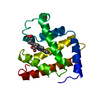 2mbwS S: Starting model for refinement |
|---|---|
| Similar structure data |
-Links
 PDBj
PDBj


- Assembly
| Deposited unit | 
| ||||||||||||
|---|---|---|---|---|---|---|---|---|---|---|---|---|---|
| 1 |
| ||||||||||||
| Unit cell |
|
-Components
-Protein , 1 types, 1 molecules A
| #1: Protein | Mass: 17355.145 Da / Num. of mol.: 1 / Mutation: H64Q Source method: isolated from a genetically manipulated source Source: (gene. exp.)  |
|---|
-Non-polymers , 5 types, 193 molecules 


| #2: Chemical | ChemComp-HEM /  Mass: 616.487 Da / Num. of mol.: 1 / Source method: obtained synthetically / Formula: C34H32FeN4O4 Mass: 616.487 Da / Num. of mol.: 1 / Source method: obtained synthetically / Formula: C34H32FeN4O4 | ||||
|---|---|---|---|---|---|
| #3: Chemical | ChemComp-ACM /  Mass: 59.067 Da / Num. of mol.: 1 / Source method: obtained synthetically / Formula: C2H5NO / Feature type: SUBJECT OF INVESTIGATION Mass: 59.067 Da / Num. of mol.: 1 / Source method: obtained synthetically / Formula: C2H5NO / Feature type: SUBJECT OF INVESTIGATION | ||||
| #4: Chemical |  Mass: 92.094 Da / Num. of mol.: 2 / Source method: obtained synthetically / Formula: C3H8O3 Mass: 92.094 Da / Num. of mol.: 2 / Source method: obtained synthetically / Formula: C3H8O3#5: Chemical | ChemComp-SO4 /  Mass: 96.063 Da / Num. of mol.: 4 / Source method: obtained synthetically / Formula: SO4 Mass: 96.063 Da / Num. of mol.: 4 / Source method: obtained synthetically / Formula: SO4#6: Water | ChemComp-HOH / | Mass: 18.015 Da / Num. of mol.: 185 / Source method: isolated from a natural source / Formula: H2O |
-Details
| Has ligand of interest | Y |
|---|
-Experimental details
-Experiment
| Experiment | Method: X-RAY DIFFRACTION / Number of used crystals: 1 |
|---|
- Sample preparation
| Crystal | Density Matthews: 3.07 Å3/Da / Density % sol: 59.91 % |
|---|---|
| Crystal grow | Temperature: 295 K / Method: vapor diffusion, hanging drop Details: 2.56-2.76 M ammonium sulfate, 100 mM Tris-HCl, 1 mM EDTA, pH 7.4 or 9.0 |
-Data collection
| Diffraction | Mean temperature: 100 K / Serial crystal experiment: N |
|---|---|
| Diffraction source | Source: ROTATING ANODE / Type: RIGAKU MICROMAX-007 HF / Wavelength: 1.5418 Å |
| Detector | Type: DECTRIS PILATUS 300K / Detector: PIXEL / Date: Feb 23, 2018 |
| Radiation | Protocol: SINGLE WAVELENGTH / Monochromatic (M) / Laue (L): M / Scattering type: x-ray |
| Radiation wavelength | Wavelength: 1.5418 Å / Relative weight: 1 |
| Reflection | Resolution: 1.76→45.26 Å / Num. obs: 21044 / % possible obs: 99.8 % / Redundancy: 1.1 % / Biso Wilson estimate: 11.64 Å2 / CC1/2: 0.999 / Rmerge(I) obs: 0.06 / Net I/σ(I): 34.32 |
| Reflection shell | Resolution: 1.76→1.823 Å / Rmerge(I) obs: 0.224 / Num. unique obs: 2046 / CC1/2: 0.999 / % possible all: 98.32 |
- Processing
| Software |
| |||||||||||||||||||||||||||||||||||||||||||||||||||||||||||||||
|---|---|---|---|---|---|---|---|---|---|---|---|---|---|---|---|---|---|---|---|---|---|---|---|---|---|---|---|---|---|---|---|---|---|---|---|---|---|---|---|---|---|---|---|---|---|---|---|---|---|---|---|---|---|---|---|---|---|---|---|---|---|---|---|---|
| Refinement | Method to determine structure: MOLECULAR REPLACEMENT Starting model: PDB entry 2MBW Resolution: 1.76→45.26 Å / SU ML: 0.1489 / Cross valid method: FREE R-VALUE / σ(F): 1.34 / Phase error: 19.5527 Stereochemistry target values: GeoStd + Monomer Library + CDL v1.2
| |||||||||||||||||||||||||||||||||||||||||||||||||||||||||||||||
| Solvent computation | Shrinkage radii: 0.9 Å / VDW probe radii: 1.11 Å / Solvent model: FLAT BULK SOLVENT MODEL | |||||||||||||||||||||||||||||||||||||||||||||||||||||||||||||||
| Displacement parameters | Biso mean: 13.75 Å2 | |||||||||||||||||||||||||||||||||||||||||||||||||||||||||||||||
| Refinement step | Cycle: LAST / Resolution: 1.76→45.26 Å
| |||||||||||||||||||||||||||||||||||||||||||||||||||||||||||||||
| Refine LS restraints |
| |||||||||||||||||||||||||||||||||||||||||||||||||||||||||||||||
| LS refinement shell |
|
