+ Open data
- Basic information
| Entry | Database: PDB / ID: 8fai | ||||||||||||
|---|---|---|---|---|---|---|---|---|---|---|---|---|---|
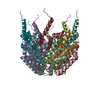
| Title | Cryo-EM structure of the Agrobacterium T-pilus | ||||||||||||
 Components Components | Protein virB2 | ||||||||||||
 Keywords Keywords | PROTEIN FIBRIL / VirB/D4 T4SS / Bacterial conjugation / T-pilus / helical filament | ||||||||||||
| Function / homology | Conjugal transfer TrbC/type IV secretion VirB2 / TrbC/VIRB2 pilin / type IV secretion system complex / protein secretion by the type IV secretion system / cell outer membrane / : / Protein virB2 Function and homology information Function and homology information | ||||||||||||
| Biological species |  Agrobacterium tumefaciens (bacteria) Agrobacterium tumefaciens (bacteria) | ||||||||||||
| Method | ELECTRON MICROSCOPY / helical reconstruction / cryo EM / Resolution: 3 Å | ||||||||||||
 Authors Authors | Kreida, S. / Narita, A. / Johnson, M.D. / Tocheva, E.I. / Das, A. / Jensen, G.J. / Ghosal, D. | ||||||||||||
| Funding support |  United States, United States,  Australia, Australia,  Canada, 3items Canada, 3items
| ||||||||||||
 Citation Citation |  Journal: Structure / Year: 2023 Journal: Structure / Year: 2023Title: Cryo-EM structure of the Agrobacterium tumefaciens T4SS-associated T-pilus reveals stoichiometric protein-phospholipid assembly. Authors: Stefan Kreida / Akihiro Narita / Matthew D Johnson / Elitza I Tocheva / Anath Das / Debnath Ghosal / Grant J Jensen /      Abstract: Agrobacterium tumefaciens causes crown gall disease in plants by the horizontal transfer of oncogenic DNA. The conjugation is mediated by the VirB/D4 type 4 secretion system (T4SS) that assembles an ...Agrobacterium tumefaciens causes crown gall disease in plants by the horizontal transfer of oncogenic DNA. The conjugation is mediated by the VirB/D4 type 4 secretion system (T4SS) that assembles an extracellular filament, the T-pilus, and is involved in mating pair formation between A. tumefaciens and the recipient plant cell. Here, we present a 3 Å cryoelectron microscopy (cryo-EM) structure of the T-pilus solved by helical reconstruction. Our structure reveals that the T-pilus is a stoichiometric assembly of the VirB2 major pilin and phosphatidylglycerol (PG) phospholipid with 5-start helical symmetry. We show that PG head groups and the positively charged Arg 91 residues of VirB2 protomers form extensive electrostatic interactions in the lumen of the T-pilus. Mutagenesis of Arg 91 abolished pilus formation. While our T-pilus structure is architecturally similar to previously published conjugative pili structures, the T-pilus lumen is narrower and positively charged, raising questions of whether the T-pilus is a conduit for ssDNA transfer. | ||||||||||||
| History |
|
- Structure visualization
| Structure viewer | Molecule:  Molmil Molmil Jmol/JSmol Jmol/JSmol |
|---|
- Downloads & links
- Download
| PDBx/mmCIF format |  8fai.cif.gz 8fai.cif.gz | 184.2 KB | Display |  PDBx/mmCIF format PDBx/mmCIF format |
|---|---|---|---|---|
| PDB format |  pdb8fai.ent.gz pdb8fai.ent.gz | 148.7 KB | Display |  PDB format PDB format |
| PDBx/mmJSON format |  8fai.json.gz 8fai.json.gz | Tree view |  PDBx/mmJSON format PDBx/mmJSON format | |
| Others |  Other downloads Other downloads |
-Validation report
| Summary document |  8fai_validation.pdf.gz 8fai_validation.pdf.gz | 1.9 MB | Display |  wwPDB validaton report wwPDB validaton report |
|---|---|---|---|---|
| Full document |  8fai_full_validation.pdf.gz 8fai_full_validation.pdf.gz | 2 MB | Display | |
| Data in XML |  8fai_validation.xml.gz 8fai_validation.xml.gz | 39.9 KB | Display | |
| Data in CIF |  8fai_validation.cif.gz 8fai_validation.cif.gz | 59.7 KB | Display | |
| Arichive directory |  https://data.pdbj.org/pub/pdb/validation_reports/fa/8fai https://data.pdbj.org/pub/pdb/validation_reports/fa/8fai ftp://data.pdbj.org/pub/pdb/validation_reports/fa/8fai ftp://data.pdbj.org/pub/pdb/validation_reports/fa/8fai | HTTPS FTP |
-Related structure data
| Related structure data |  28957MC M: map data used to model this data C: citing same article ( |
|---|---|
| Similar structure data | Similarity search - Function & homology  F&H Search F&H Search |
- Links
Links
- Assembly
| Deposited unit | 
|
|---|---|
| 1 |
|
- Components
| #1: Protein | Mass: 12326.439 Da / Num. of mol.: 15 / Source method: isolated from a natural source / Source: (natural)  Agrobacterium tumefaciens (bacteria) / Strain: NT1REB(pJK270) / References: UniProt: P17792 Agrobacterium tumefaciens (bacteria) / Strain: NT1REB(pJK270) / References: UniProt: P17792#2: Chemical | ChemComp-XL0 / ( Mass: 746.991 Da / Num. of mol.: 15 / Source method: obtained synthetically / Formula: C40H75O10P / Feature type: SUBJECT OF INVESTIGATION Has ligand of interest | Y | |
|---|
-Experimental details
-Experiment
| Experiment | Method: ELECTRON MICROSCOPY |
|---|---|
| EM experiment | Aggregation state: FILAMENT / 3D reconstruction method: helical reconstruction |
- Sample preparation
| Component |
| ||||||||||||||||||||||||
|---|---|---|---|---|---|---|---|---|---|---|---|---|---|---|---|---|---|---|---|---|---|---|---|---|---|
| Molecular weight |
| ||||||||||||||||||||||||
| Source (natural) |
| ||||||||||||||||||||||||
| Buffer solution | pH: 5.5 | ||||||||||||||||||||||||
| Buffer component | Conc.: 50 mM / Name: 2-(N-morpholino)ethanesulfonic acid / Formula: MES | ||||||||||||||||||||||||
| Specimen | Conc.: 1 mg/ml / Embedding applied: NO / Shadowing applied: NO / Staining applied: NO / Vitrification applied: YES | ||||||||||||||||||||||||
| Specimen support | Grid material: GOLD / Grid mesh size: 300 divisions/in. / Grid type: UltrAuFoil R1.2/1.3 | ||||||||||||||||||||||||
| Vitrification | Instrument: FEI VITROBOT MARK IV / Cryogen name: ETHANE-PROPANE / Humidity: 100 % / Chamber temperature: 295 K |
- Electron microscopy imaging
| Experimental equipment |  Model: Titan Krios / Image courtesy: FEI Company |
|---|---|
| Microscopy | Model: FEI TITAN KRIOS |
| Electron gun | Electron source:  FIELD EMISSION GUN / Accelerating voltage: 300 kV / Illumination mode: FLOOD BEAM FIELD EMISSION GUN / Accelerating voltage: 300 kV / Illumination mode: FLOOD BEAM |
| Electron lens | Mode: BRIGHT FIELD / Nominal magnification: 105000 X / Nominal defocus max: 2000 nm / Nominal defocus min: 700 nm / Cs: 2.7 mm / C2 aperture diameter: 100 µm |
| Image recording | Electron dose: 60 e/Å2 / Film or detector model: GATAN K3 (6k x 4k) |
- Processing
| Software | Name: PHENIX / Version: 1.19.2_4158: / Classification: refinement | ||||||||||||||||||||||||||||||||||||||||
|---|---|---|---|---|---|---|---|---|---|---|---|---|---|---|---|---|---|---|---|---|---|---|---|---|---|---|---|---|---|---|---|---|---|---|---|---|---|---|---|---|---|
| EM software |
| ||||||||||||||||||||||||||||||||||||||||
| CTF correction | Type: PHASE FLIPPING AND AMPLITUDE CORRECTION | ||||||||||||||||||||||||||||||||||||||||
| Helical symmerty |
| ||||||||||||||||||||||||||||||||||||||||
| Particle selection | Num. of particles selected: 10934 Details: Filaments were manually picked using EMAN2 e2helixboxer and particles were extracted by RELION. | ||||||||||||||||||||||||||||||||||||||||
| 3D reconstruction | Resolution: 3 Å / Resolution method: FSC 0.143 CUT-OFF / Num. of particles: 3543 / Num. of class averages: 2 / Symmetry type: HELICAL | ||||||||||||||||||||||||||||||||||||||||
| Refine LS restraints |
|
 Movie
Movie Controller


 PDBj
PDBj