 Movie
Movie Controller

[English] 日本語
 Yorodumi
- PDB-8fa1: Cryo-EM structure of the SARS-CoV-2 HR1HR2 fusion core complex wi... -
+ Open data

- Basic information
| Entry | Database: PDB / ID: 8fa1 | ||||||
|---|---|---|---|---|---|---|---|
| Title | Cryo-EM structure of the SARS-CoV-2 HR1HR2 fusion core complex with N969K mutation | ||||||
 Components Components |
| ||||||
 Keywords Keywords | VIRAL PROTEIN / spike / HR1HR2 / fusion / scaffold | ||||||
| Function / homology |  Function and homology information Function and homology informationoxidoreductase activity, acting on metal ions / ferric iron binding / symbiont-mediated disruption of host tissue / Maturation of spike protein / Translation of Structural Proteins / Virion Assembly and Release / host cell surface / host extracellular region / symbiont-mediated-mediated suppression of host tetherin activity / Induction of Cell-Cell Fusion ...oxidoreductase activity, acting on metal ions / ferric iron binding / symbiont-mediated disruption of host tissue / Maturation of spike protein / Translation of Structural Proteins / Virion Assembly and Release / host cell surface / host extracellular region / symbiont-mediated-mediated suppression of host tetherin activity / Induction of Cell-Cell Fusion / structural constituent of virion / positive regulation of viral entry into host cell / membrane fusion / host cell endoplasmic reticulum-Golgi intermediate compartment membrane / Attachment and Entry / entry receptor-mediated virion attachment to host cell / receptor-mediated virion attachment to host cell / host cell surface receptor binding / symbiont-mediated suppression of host innate immune response / endocytosis involved in viral entry into host cell / receptor ligand activity / fusion of virus membrane with host plasma membrane / fusion of virus membrane with host endosome membrane / viral envelope / symbiont entry into host cell / virion attachment to host cell / host cell plasma membrane / SARS-CoV-2 activates/modulates innate and adaptive immune responses / virion membrane / membrane / identical protein binding / plasma membrane Similarity search - Function | ||||||
| Biological species |  Nostoc punctiforme PCC 73102 (bacteria) Nostoc punctiforme PCC 73102 (bacteria)  Severe acute respiratory syndrome coronavirus 2 Severe acute respiratory syndrome coronavirus 2 | ||||||
| Method | ELECTRON MICROSCOPY / single particle reconstruction / cryo EM / Resolution: 2.51 Å | ||||||
 Authors Authors | Yang, K. / Brunger, A.T. | ||||||
| Funding support |  United States, 1items United States, 1items
| ||||||
 Citation Citation | Journal: Proc Natl Acad Sci U S A / Year: 2023 Title: Structure-based design of a SARS-CoV-2 Omicron-specific inhibitor. Authors: Kailu Yang / Chuchu Wang / Alex J B Kreutzberger / K Ian White / Richard A Pfuetzner / Luis Esquivies / Tomas Kirchhausen / Axel T Brunger / Abstract: The Omicron variant of severe acute respiratory syndrome coronavirus 2 (SARS-CoV-2) introduced a relatively large number of mutations, including three mutations in the highly conserved heptad repeat ...The Omicron variant of severe acute respiratory syndrome coronavirus 2 (SARS-CoV-2) introduced a relatively large number of mutations, including three mutations in the highly conserved heptad repeat 1 (HR1) region of the spike glycoprotein (S) critical for its membrane fusion activity. We show that one of these mutations, N969K induces a substantial displacement in the structure of the heptad repeat 2 (HR2) backbone in the HR1HR2 postfusion bundle. Due to this mutation, fusion-entry peptide inhibitors based on the Wuhan strain sequence are less efficacious. Here, we report an Omicron-specific peptide inhibitor designed based on the structure of the Omicron HR1HR2 postfusion bundle. Specifically, we inserted an additional residue in HR2 near the Omicron HR1 K969 residue to better accommodate the N969K mutation and relieve the distortion in the structure of the HR1HR2 postfusion bundle it introduced. The designed inhibitor recovers the loss of inhibition activity of the original longHR2_42 peptide with the Wuhan strain sequence against the Omicron variant in both a cell-cell fusion assay and a vesicular stomatitis virus (VSV)-SARS-CoV-2 chimera infection assay, suggesting that a similar approach could be used to combat future variants. From a mechanistic perspective, our work suggests the interactions in the extended region of HR2 may mediate the initial landing of HR2 onto HR1 during the transition of the S protein from the prehairpin intermediate to the postfusion state. | ||||||
| History |
|
- Structure visualization
| Structure viewer | Molecule: MolmilJmol/JSmol |
|---|
- Downloads & links
-Download
| PDBx/mmCIF format | 8fa1.cif.gz | 79.9 KB | Display | PDBx/mmCIF format |
|---|---|---|---|---|
| PDB format | pdb8fa1.ent.gz | 56.3 KB | Display | PDB format |
| PDBx/mmJSON format | 8fa1.json.gz | Tree view | PDBx/mmJSON format | |
| Others |  Other downloads Other downloads |
-Validation report
| Arichive directory | https://data.pdbj.org/pub/pdb/validation_reports/fa/8fa1ftp://data.pdbj.org/pub/pdb/validation_reports/fa/8fa1 | HTTPS FTP |
|---|
-Related structure data
| Related structure data | 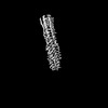 28947MC  7tikC  8fa2C M: map data used to model this data C: citing same article ( |
|---|---|
| Similar structure data |


-Links
 PDBj
PDBj


- Assembly
| Deposited unit | 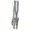
|
|---|---|
| 1 |
|
-Components
| #1: Protein | Mass: 28774.133 Da / Num. of mol.: 3 Source method: isolated from a genetically manipulated source Source: (gene. exp.) Nostoc punctiforme PCC 73102 (bacteria), (gene. exp.) Severe acute respiratory syndrome coronavirus 2Gene: Npun_R5799 / Production host: #2: Protein/peptide | Mass: 4935.439 Da / Num. of mol.: 3 Source method: isolated from a genetically manipulated source Source: (gene. exp.) Severe acute respiratory syndrome coronavirus 2Gene: S, 2 / Production host: |
|---|
-Experimental details
-Experiment
| Experiment | Method: ELECTRON MICROSCOPY |
|---|---|
| EM experiment | Aggregation state: PARTICLE / 3D reconstruction method: single particle reconstruction |
- Sample preparation
| Component | Name: SARS-CoV-2 HR1HR2 complex / Type: COMPLEX / Entity ID: all / Source: RECOMBINANT |
|---|---|
| Molecular weight | Value: 0.04 MDa / Experimental value: NO |
| Source (natural) | Organism: Severe acute respiratory syndrome coronavirus 2 |
| Source (recombinant) | Organism: |
| Buffer solution | pH: 7.4 |
| Specimen | Embedding applied: NO / Shadowing applied: NO / Staining applied: NO / Vitrification applied: YES |
| Vitrification | Cryogen name: ETHANE |
- Electron microscopy imaging
| Experimental equipment |  Model: Titan Krios / Image courtesy: FEI Company |
|---|---|
| Microscopy | Model: FEI TITAN KRIOS |
| Electron gun | Electron source:  FIELD EMISSION GUN / Accelerating voltage: 300 kV / Illumination mode: FLOOD BEAM FIELD EMISSION GUN / Accelerating voltage: 300 kV / Illumination mode: FLOOD BEAM |
| Electron lens | Mode: BRIGHT FIELD / Nominal defocus max: 2000 nm / Nominal defocus min: 300 nm |
| Image recording | Electron dose: 55 e/Å2 / Film or detector model: GATAN K3 (6k x 4k) |
- Processing
| CTF correction | Type: NONE |
|---|---|
| 3D reconstruction | Resolution: 2.51 Å / Resolution method: FSC 0.143 CUT-OFF / Num. of particles: 883152 / Symmetry type: POINT |
