 Movie
Movie Controller


+ Open data

- Basic information
| Entry | Database: PDB / ID: 8f91 | |||||||||
|---|---|---|---|---|---|---|---|---|---|---|
| Title | OxyB, a cytochrome P450 involved in keratinimicin biosynthesis | |||||||||
 Components Components | OxyB | |||||||||
 Keywords Keywords | OXIDOREDUCTASE / Cytochrome P450 / monooxygenase / keratinimicin | |||||||||
| Function / homology |  Function and homology information Function and homology informationoxidoreductase activity, acting on paired donors, with incorporation or reduction of molecular oxygen / monooxygenase activity / iron ion binding / heme binding Similarity search - Function | |||||||||
| Biological species |  Amycolatopsis keratiniphila (bacteria) Amycolatopsis keratiniphila (bacteria) | |||||||||
| Method |  X-RAY DIFFRACTION / SYNCHROTRON / MOLECULAR REPLACEMENT / Resolution: 2.8 Å X-RAY DIFFRACTION / SYNCHROTRON / MOLECULAR REPLACEMENT / Resolution: 2.8 Å | |||||||||
 Authors Authors | Ireland, K.A. / Davis, K.M. | |||||||||
| Funding support |  United States, 2items United States, 2items
| |||||||||
 Citation Citation | Journal: Acs Chem.Biol. / Year: 2023 Title: Robust Chemoenzymatic Synthesis of Keratinimicin Aglycone Analogues Facilitated by the Structure and Selectivity of OxyB. Authors: Hauser, N. / Ireland, K.A. / Chioti, V.T. / Forneris, C.C. / Davis, K.M. / Seyedsayamdost, M.R. | |||||||||
| History |
|
- Structure visualization
| Structure viewer | Molecule: MolmilJmol/JSmol |
|---|
- Downloads & links
-Download
| PDBx/mmCIF format | 8f91.cif.gz | 92.7 KB | Display | PDBx/mmCIF format |
|---|---|---|---|---|
| PDB format | pdb8f91.ent.gz | 60.4 KB | Display | PDB format |
| PDBx/mmJSON format | 8f91.json.gz | Tree view | PDBx/mmJSON format | |
| Others |  Other downloads Other downloads |
-Validation report
| Summary document | 8f91_validation.pdf.gz | 785.9 KB | Display | wwPDB validaton report |
|---|---|---|---|---|
| Full document | 8f91_full_validation.pdf.gz | 791.3 KB | Display | |
| Data in XML | 8f91_validation.xml.gz | 15.6 KB | Display | |
| Data in CIF | 8f91_validation.cif.gz | 20.4 KB | Display | |
| Arichive directory | https://data.pdbj.org/pub/pdb/validation_reports/f9/8f91ftp://data.pdbj.org/pub/pdb/validation_reports/f9/8f91 | HTTPS FTP |
-Related structure data
| Related structure data |  5ex6S S: Starting model for refinement |
|---|---|
| Similar structure data |
-Links
 PDBj
PDBj
- Assembly
| Deposited unit | 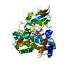
| ||||||||||||
|---|---|---|---|---|---|---|---|---|---|---|---|---|---|
| 1 |
| ||||||||||||
| Unit cell |
|
-Components
| #1: Protein | Mass: 46425.711 Da / Num. of mol.: 1 Source method: isolated from a genetically manipulated source Source: (gene. exp.) Amycolatopsis keratiniphila (bacteria) / Strain: NRRL B24117 / Production host: |
|---|---|
| #2: Chemical | ChemComp-HEM /   Mass: 616.487 Da / Num. of mol.: 1 / Source method: obtained synthetically / Formula: C34H32FeN4O4 Mass: 616.487 Da / Num. of mol.: 1 / Source method: obtained synthetically / Formula: C34H32FeN4O4 |
| #3: Water | ChemComp-HOH /  Mass: 18.015 Da / Num. of mol.: 19 / Source method: isolated from a natural source / Formula: H2O Mass: 18.015 Da / Num. of mol.: 19 / Source method: isolated from a natural source / Formula: H2O |
| Has ligand of interest | N |
-Experimental details
-Experiment
| Experiment | Method: X-RAY DIFFRACTION / Number of used crystals: 1 |
|---|
- Sample preparation
| Crystal | Density Matthews: 2.54 Å3/Da / Density % sol: 51.5 % |
|---|---|
| Crystal grow | Temperature: 293 K / Method: vapor diffusion, sitting drop / pH: 7 / Details: 1.8 ammonium citrate tribasic, pH 7 / Temp details: Room temperature |
-Data collection
| Diffraction | Mean temperature: 100 K / Serial crystal experiment: N |
|---|---|
| Diffraction source | Source: SYNCHROTRON / Site: APS / Beamline: 21-ID-F / Wavelength: 0.9787 Å |
| Detector | Type: RAYONIX MX-300 / Detector: CCD / Date: Feb 6, 2022 |
| Radiation | Monochromator: C(111) / Protocol: SINGLE WAVELENGTH / Monochromatic (M) / Laue (L): M / Scattering type: x-ray |
| Radiation wavelength | Wavelength: 0.9787 Å / Relative weight: 1 |
| Reflection | Resolution: 2.8→56.62 Å / Num. obs: 12403 / % possible obs: 99.95 % / Redundancy: 2 % / Biso Wilson estimate: 67.47 Å2 / CC1/2: 1 / CC star: 1 / Rmerge(I) obs: 0.0177 / Rpim(I) all: 0.0177 / Rrim(I) all: 0.02503 / Net I/σ(I): 20.95 |
| Reflection shell | Resolution: 2.8→2.9 Å / Redundancy: 2 % / Rmerge(I) obs: 0.2136 / Mean I/σ(I) obs: 3.21 / Num. unique obs: 1208 / CC1/2: 0.829 / CC star: 0.952 / Rpim(I) all: 0.2136 / Rrim(I) all: 0.302 / % possible all: 100 |
- Processing
| Software |
| |||||||||||||||||||||||||||||||||||
|---|---|---|---|---|---|---|---|---|---|---|---|---|---|---|---|---|---|---|---|---|---|---|---|---|---|---|---|---|---|---|---|---|---|---|---|---|
| Refinement | Method to determine structure: MOLECULAR REPLACEMENT Starting model: PDB entry 5EX6 Resolution: 2.8→56.62 Å / SU ML: 0.3824 / Cross valid method: FREE R-VALUE / σ(F): 1.34 / Phase error: 29.1207 Stereochemistry target values: GeoStd + Monomer Library + CDL v1.2
| |||||||||||||||||||||||||||||||||||
| Solvent computation | Shrinkage radii: 0.9 Å / VDW probe radii: 1.1 Å / Solvent model: FLAT BULK SOLVENT MODEL | |||||||||||||||||||||||||||||||||||
| Displacement parameters | Biso mean: 68.45 Å2 | |||||||||||||||||||||||||||||||||||
| Refinement step | Cycle: LAST / Resolution: 2.8→56.62 Å
| |||||||||||||||||||||||||||||||||||
| Refine LS restraints |
| |||||||||||||||||||||||||||||||||||
| LS refinement shell |
|
