 Movie
Movie Controller


+ Open data

- Basic information
| Entry | Database: PDB / ID: 8f7n | |||||||||
|---|---|---|---|---|---|---|---|---|---|---|
| Title | Crystal structure of chemoreceptor McpZ ligand sensing domain | |||||||||
 Components Components | Methyl-accepting chemotaxis protein | |||||||||
 Keywords Keywords | SIGNALING PROTEIN / MCP (Methyl-accepting chemotaxis protein) / Chemotaxis / Chemoreceptor | |||||||||
| Function / homology |  Function and homology information Function and homology information | |||||||||
| Biological species |  Sinorhizobium meliloti (bacteria) Sinorhizobium meliloti (bacteria) | |||||||||
| Method |  X-RAY DIFFRACTION / SYNCHROTRON / SAD / Resolution: 2.7 Å X-RAY DIFFRACTION / SYNCHROTRON / SAD / Resolution: 2.7 Å | |||||||||
 Authors Authors | Salar, S. / Schubot, F.D. | |||||||||
| Funding support |  United States, 2items United States, 2items
| |||||||||
 Citation Citation | Journal: Proteins / Year: 2023 Title: The structural analysis of the periplasmic domain of Sinorhizobium meliloti chemoreceptor McpZ reveals a novel fold and suggests a complex mechanism of transmembrane signaling. Authors: Salar, S. / Ball, N.E. / Baaziz, H. / Nix, J.C. / Sobe, R.C. / Compton, K.K. / Zhulin, I.B. / Brown, A.M. / Scharf, B.E. / Schubot, F.D. | |||||||||
| History |
|
- Structure visualization
| Structure viewer | Molecule: MolmilJmol/JSmol |
|---|
- Downloads & links
-Download
| PDBx/mmCIF format | 8f7n.cif.gz | 218 KB | Display | PDBx/mmCIF format |
|---|---|---|---|---|
| PDB format | pdb8f7n.ent.gz | 177.2 KB | Display | PDB format |
| PDBx/mmJSON format | 8f7n.json.gz | Tree view | PDBx/mmJSON format | |
| Others |  Other downloads Other downloads |
-Validation report
| Summary document | 8f7n_validation.pdf.gz | 436.5 KB | Display | wwPDB validaton report |
|---|---|---|---|---|
| Full document | 8f7n_full_validation.pdf.gz | 450.1 KB | Display | |
| Data in XML | 8f7n_validation.xml.gz | 16.3 KB | Display | |
| Data in CIF | 8f7n_validation.cif.gz | 21.6 KB | Display | |
| Arichive directory | https://data.pdbj.org/pub/pdb/validation_reports/f7/8f7nftp://data.pdbj.org/pub/pdb/validation_reports/f7/8f7n | HTTPS FTP |
-Related structure data
| Similar structure data |
|---|
-Links
 PDBj
PDBj


- Assembly
| Deposited unit | 
| ||||||||
|---|---|---|---|---|---|---|---|---|---|
| 1 | 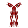
| ||||||||
| Unit cell |
| ||||||||
| Components on special symmetry positions |
|
-Components
| #1: Protein | Mass: 41802.164 Da / Num. of mol.: 1 Source method: isolated from a genetically manipulated source Source: (gene. exp.) Sinorhizobium meliloti (bacteria) / Gene: CN211_06960 / Production host: | ||||
|---|---|---|---|---|---|
| #2: Chemical | ChemComp-YB /   Mass: 173.040 Da / Num. of mol.: 10 / Source method: obtained synthetically / Formula: Yb Mass: 173.040 Da / Num. of mol.: 10 / Source method: obtained synthetically / Formula: Yb#3: Water | ChemComp-HOH / |  Mass: 18.015 Da / Num. of mol.: 9 / Source method: isolated from a natural source / Formula: H2O Mass: 18.015 Da / Num. of mol.: 9 / Source method: isolated from a natural source / Formula: H2OHas ligand of interest | N | |
-Experimental details
-Experiment
| Experiment | Method: X-RAY DIFFRACTION / Number of used crystals: 1 |
|---|
- Sample preparation
| Crystal grow | Temperature: 295.15 K / Method: vapor diffusion, sitting drop Details: Lanthanides, MOPSO, Bis-Tris pH 6.5, and Precipitant Mix 8 (PEG 20000, Trimethylpropane, NDSB 195) |
|---|
-Data collection
| Diffraction | Mean temperature: 100 K / Serial crystal experiment: N |
|---|---|
| Diffraction source | Source: SYNCHROTRON / Site: ALS / Beamline: 4.2.2 / Wavelength: 1 Å |
| Detector | Type: RDI CMOS_8M / Detector: CMOS / Date: Jul 10, 2021 |
| Radiation | Protocol: SINGLE WAVELENGTH / Monochromatic (M) / Laue (L): M / Scattering type: x-ray |
| Radiation wavelength | Wavelength: 1 Å / Relative weight: 1 |
| Reflection | Resolution: 2.54→46.6 Å / Num. obs: 32346 / % possible obs: 99.9 % / Redundancy: 20 % / Biso Wilson estimate: 75.86 Å2 / CC1/2: 1 / Rmerge(I) obs: 0.166 / Rpim(I) all: 0.031 / Rrim(I) all: 0.169 / Χ2: 0.84 / Net I/σ(I): 17.8 |
| Reflection shell | Resolution: 2.54→2.65 Å / Mean I/σ(I) obs: 0.5 / Num. unique obs: 3873 / CC1/2: 0.41 / Χ2: 0.64 / % possible all: 100 |
- Processing
| Software |
| |||||||||||||||||||||||||||||||||||||||||||||||||||||||||||||||||||||||||||||||||||||||||||||||||||||||||||||||||||||||||||||||||||||
|---|---|---|---|---|---|---|---|---|---|---|---|---|---|---|---|---|---|---|---|---|---|---|---|---|---|---|---|---|---|---|---|---|---|---|---|---|---|---|---|---|---|---|---|---|---|---|---|---|---|---|---|---|---|---|---|---|---|---|---|---|---|---|---|---|---|---|---|---|---|---|---|---|---|---|---|---|---|---|---|---|---|---|---|---|---|---|---|---|---|---|---|---|---|---|---|---|---|---|---|---|---|---|---|---|---|---|---|---|---|---|---|---|---|---|---|---|---|---|---|---|---|---|---|---|---|---|---|---|---|---|---|---|---|---|
| Refinement | Method to determine structure: SAD / Resolution: 2.7→46.6 Å / SU ML: 0.34 / Cross valid method: FREE R-VALUE / σ(F): 1.34 / Phase error: 32.51 / Stereochemistry target values: ML
| |||||||||||||||||||||||||||||||||||||||||||||||||||||||||||||||||||||||||||||||||||||||||||||||||||||||||||||||||||||||||||||||||||||
| Solvent computation | Shrinkage radii: 0.9 Å / VDW probe radii: 1.11 Å / Solvent model: FLAT BULK SOLVENT MODEL | |||||||||||||||||||||||||||||||||||||||||||||||||||||||||||||||||||||||||||||||||||||||||||||||||||||||||||||||||||||||||||||||||||||
| Refinement step | Cycle: LAST / Resolution: 2.7→46.6 Å
| |||||||||||||||||||||||||||||||||||||||||||||||||||||||||||||||||||||||||||||||||||||||||||||||||||||||||||||||||||||||||||||||||||||
| Refine LS restraints |
| |||||||||||||||||||||||||||||||||||||||||||||||||||||||||||||||||||||||||||||||||||||||||||||||||||||||||||||||||||||||||||||||||||||
| LS refinement shell |
| |||||||||||||||||||||||||||||||||||||||||||||||||||||||||||||||||||||||||||||||||||||||||||||||||||||||||||||||||||||||||||||||||||||
| Refinement TLS params. | Method: refined / Origin x: 75.1245 Å / Origin y: 45.3564 Å / Origin z: 16.535 Å
| |||||||||||||||||||||||||||||||||||||||||||||||||||||||||||||||||||||||||||||||||||||||||||||||||||||||||||||||||||||||||||||||||||||
| Refinement TLS group | Selection details: all |
