NURF complex / endoderm development / anterior/posterior pattern specification / ATPase complex / : / embryonic placenta development / cellular response to nerve growth factor stimulus / brain development / cell body / sequence-specific DNA binding ...NURF complex / endoderm development / anterior/posterior pattern specification / ATPase complex / : / embryonic placenta development / cellular response to nerve growth factor stimulus / brain development / cell body / sequence-specific DNA binding / RNA polymerase II cis-regulatory region sequence-specific DNA binding / chromatin remodeling / dendrite / regulation of DNA-templated transcription / regulation of transcription by RNA polymerase II / chromatin / perinuclear region of cytoplasm / negative regulation of transcription by RNA polymerase II / positive regulation of transcription by RNA polymerase II / extracellular exosome / zinc ion binding / nucleoplasm / nucleus / cytoplasm Similarity search - Function
Nucleosome-remodelingfactorsubunitBPTF / Bromodomain and PHD finger-containing transcription factor / Fetal Alz-50 clone 1 protein / Fetal ...Bromodomain and PHD finger-containing transcription factor / Fetal Alz-50 clone 1 protein / Fetal Alzheimer antigen
Mass: 14455.415 Da / Num. of mol.: 1 / Fragment: BPTF bromodomain (uniprot residues 2917-3037) Source method: isolated from a genetically manipulated source Details: The following residues were not located. SER A 2915 MET A 2916 Source: (gene. exp.) Homo sapiens (human) / Gene: BPTF, FAC1, FALZ / Production host: Escherichia coli (E. coli) / References: UniProt: Q12830
In the structure databanks used in Yorodumi, some data are registered as the other names, "COVID-19 virus" and "2019-nCoV". Here are the details of the virus and the list of structure data.
Jan 31, 2019. EMDB accession codes are about to change! (news from PDBe EMDB page)
EMDB accession codes are about to change! (news from PDBe EMDB page)
The allocation of 4 digits for EMDB accession codes will soon come to an end. Whilst these codes will remain in use, new EMDB accession codes will include an additional digit and will expand incrementally as the available range of codes is exhausted. The current 4-digit format prefixed with “EMD-” (i.e. EMD-XXXX) will advance to a 5-digit format (i.e. EMD-XXXXX), and so on. It is currently estimated that the 4-digit codes will be depleted around Spring 2019, at which point the 5-digit format will come into force.
The EM Navigator/Yorodumi systems omit the EMD- prefix.
Related info.:Q: What is EMD? / ID/Accession-code notation in Yorodumi/EM Navigator
Yorodumi is a browser for structure data from EMDB, PDB, SASBDB, etc.
This page is also the successor to EM Navigator detail page, and also detail information page/front-end page for Omokage search.
The word "yorodu" (or yorozu) is an old Japanese word meaning "ten thousand". "mi" (miru) is to see.
Related info.:EMDB / PDB / SASBDB / Comparison of 3 databanks / Yorodumi Search / Aug 31, 2016. New EM Navigator & Yorodumi / Yorodumi Papers / Jmol/JSmol / Function and homology information / Changes in new EM Navigator and Yorodumi
 Movie
Movie Controller


 Open data
 Basic information
 Components
 Keywords
 Function and homology information
 Homo sapiens (human)
 X-RAY DIFFRACTION /
  Authors
 United States, 1items
  Citation
 Structure visualization
 Downloads & links
 Other downloads
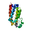
 PDBj
PDBj Assembly
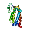

 Mass: 18.015 Da / Num. of mol.: 120 / Source method: isolated from a natural source / Formula: H2O
 Sample preparation
 Processing