 Movie
Movie Controller


+ Open data

- Basic information
| Entry | Database: PDB / ID: 8f6c | |||||||||||||||||||||||||||||||||||||||||||||||||||||||||||||||||||||||||||||||||
|---|---|---|---|---|---|---|---|---|---|---|---|---|---|---|---|---|---|---|---|---|---|---|---|---|---|---|---|---|---|---|---|---|---|---|---|---|---|---|---|---|---|---|---|---|---|---|---|---|---|---|---|---|---|---|---|---|---|---|---|---|---|---|---|---|---|---|---|---|---|---|---|---|---|---|---|---|---|---|---|---|---|---|
| Title | E. coli cytochrome bo3 ubiquinol oxidase dimer | |||||||||||||||||||||||||||||||||||||||||||||||||||||||||||||||||||||||||||||||||
 Components Components | (Cytochrome bo(3) ubiquinol oxidase subunit ...) x 4 | |||||||||||||||||||||||||||||||||||||||||||||||||||||||||||||||||||||||||||||||||
 Keywords Keywords | PROTON TRANSPORT / heme-copper oxidase / proton translocation / E. coli aerobic respiratory chain / membrane protein / Structural Genomics / Center for Structural Biology of Infectious Diseases / CSBID | |||||||||||||||||||||||||||||||||||||||||||||||||||||||||||||||||||||||||||||||||
| Function / homology |  Function and homology information Function and homology informationoxidoreduction-driven active transmembrane transporter activity / cytochrome o ubiquinol oxidase complex / ubiquinol oxidase (H+-transporting) / cytochrome bo3 ubiquinol oxidase activity / aerobic electron transport chain / respiratory chain complex / oxidoreductase activity, acting on diphenols and related substances as donors, oxygen as acceptor / cytochrome-c oxidase activity / proton transmembrane transporter activity / ubiquinone binding ...oxidoreduction-driven active transmembrane transporter activity / cytochrome o ubiquinol oxidase complex / ubiquinol oxidase (H+-transporting) / cytochrome bo3 ubiquinol oxidase activity / aerobic electron transport chain / respiratory chain complex / oxidoreductase activity, acting on diphenols and related substances as donors, oxygen as acceptor / cytochrome-c oxidase activity / proton transmembrane transporter activity / ubiquinone binding / electron transport coupled proton transport / ATP synthesis coupled electron transport / aerobic respiration / respiratory electron transport chain / electron transfer activity / copper ion binding / heme binding / plasma membrane Similarity search - Function | |||||||||||||||||||||||||||||||||||||||||||||||||||||||||||||||||||||||||||||||||
| Biological species |  | |||||||||||||||||||||||||||||||||||||||||||||||||||||||||||||||||||||||||||||||||
| Method | ELECTRON MICROSCOPY / single particle reconstruction / cryo EM / Resolution: 3.46 Å | |||||||||||||||||||||||||||||||||||||||||||||||||||||||||||||||||||||||||||||||||
 Authors Authors | Guo, Y. / Karimullina, E. / Borek, D. / Savchenko, A. / Center for Structural Biology of Infectious Diseases (CSBID) | |||||||||||||||||||||||||||||||||||||||||||||||||||||||||||||||||||||||||||||||||
| Funding support |  United States, 1items United States, 1items
| |||||||||||||||||||||||||||||||||||||||||||||||||||||||||||||||||||||||||||||||||
 Citation Citation | Journal: Protein Sci / Year: 2023 Title: Monomer and dimer structures of cytochrome bo ubiquinol oxidase from Escherichia coli. Authors: Yirui Guo / Elina Karimullina / Tabitha Emde / Zbyszek Otwinowski / Dominika Borek / Alexei Savchenko /  Abstract: The Escherichia coli cytochrome bo ubiquinol oxidase is a four-subunit heme-copper oxidase that serves as a proton pump in the E. coli aerobic respiratory chain. Despite many mechanistic studies, it ...The Escherichia coli cytochrome bo ubiquinol oxidase is a four-subunit heme-copper oxidase that serves as a proton pump in the E. coli aerobic respiratory chain. Despite many mechanistic studies, it is unclear whether this ubiquinol oxidase functions as a monomer, or as a dimer in a manner similar to its eukaryotic counterparts-the mitochondrial electron transport complexes. In this study, we determined the monomeric and dimeric structures of the E. coli cytochrome bo ubiquinol oxidase reconstituted in amphipol by cryogenic electron microscopy single particle reconstruction (cryo-EM SPR) to a resolution of 3.15 and 3.46 Å, respectively. We have discovered that the protein can form a dimer with C2 symmetry, with the dimerization interface maintained by interactions between the subunit II of one monomer and the subunit IV of the other monomer. Moreover, the dimerization does not induce significant structural changes in the monomers, except the movement of a loop in subunit IV (residues 67-74). | |||||||||||||||||||||||||||||||||||||||||||||||||||||||||||||||||||||||||||||||||
| History |
|
- Structure visualization
| Structure viewer | Molecule: MolmilJmol/JSmol |
|---|
- Downloads & links
-Download
| PDBx/mmCIF format | 8f6c.cif.gz | 450 KB | Display | PDBx/mmCIF format |
|---|---|---|---|---|
| PDB format | pdb8f6c.ent.gz | 362 KB | Display | PDB format |
| PDBx/mmJSON format | 8f6c.json.gz | Tree view | PDBx/mmJSON format | |
| Others |  Other downloads Other downloads |
-Validation report
| Arichive directory | https://data.pdbj.org/pub/pdb/validation_reports/f6/8f6cftp://data.pdbj.org/pub/pdb/validation_reports/f6/8f6c | HTTPS FTP |
|---|
-Related structure data
| Related structure data |  28879MC 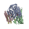 8f68C M: map data used to model this data C: citing same article ( |
|---|---|
| Similar structure data | |
| Other databases |
-Links
 PDBj
PDBj


- Assembly
| Deposited unit | 
| |||||||||||||||
|---|---|---|---|---|---|---|---|---|---|---|---|---|---|---|---|---|
| 1 |
| |||||||||||||||
| Noncrystallographic symmetry (NCS) | NCS domain:
|
-Components
-Cytochrome bo(3) ubiquinol oxidase subunit ... , 4 types, 8 molecules AEBFCGDH
| #1: Protein | Mass: 73896.875 Da / Num. of mol.: 2 Source method: isolated from a genetically manipulated source Source: (gene. exp.) References: UniProt: P0ABI8, ubiquinol oxidase (H+-transporting) #2: Protein | Mass: 28839.297 Da / Num. of mol.: 2 Source method: isolated from a genetically manipulated source Source: (gene. exp.) #3: Protein | Mass: 20464.258 Da / Num. of mol.: 2 Source method: isolated from a genetically manipulated source Source: (gene. exp.) #4: Protein | Mass: 10742.083 Da / Num. of mol.: 2 Source method: isolated from a genetically manipulated source Source: (gene. exp.) |
|---|
-Non-polymers , 4 types, 12 molecules 


| #5: Chemical |  Mass: 616.487 Da / Num. of mol.: 2 / Source method: obtained synthetically / Formula: C34H32FeN4O4 / Feature type: SUBJECT OF INVESTIGATION Mass: 616.487 Da / Num. of mol.: 2 / Source method: obtained synthetically / Formula: C34H32FeN4O4 / Feature type: SUBJECT OF INVESTIGATION#6: Chemical |  Mass: 838.854 Da / Num. of mol.: 2 / Source method: obtained synthetically / Formula: C49H58FeN4O5 / Feature type: SUBJECT OF INVESTIGATION Mass: 838.854 Da / Num. of mol.: 2 / Source method: obtained synthetically / Formula: C49H58FeN4O5 / Feature type: SUBJECT OF INVESTIGATION#7: Chemical |  Mass: 63.546 Da / Num. of mol.: 2 / Source method: obtained synthetically / Formula: Cu / Feature type: SUBJECT OF INVESTIGATION Mass: 63.546 Da / Num. of mol.: 2 / Source method: obtained synthetically / Formula: Cu / Feature type: SUBJECT OF INVESTIGATION#8: Chemical | ChemComp-3PE /  Mass: 748.065 Da / Num. of mol.: 6 / Source method: obtained synthetically / Formula: C41H82NO8P / Comment: phospholipid*YM Mass: 748.065 Da / Num. of mol.: 6 / Source method: obtained synthetically / Formula: C41H82NO8P / Comment: phospholipid*YM |
|---|
-Details
| Has ligand of interest | Y |
|---|---|
| Has protein modification | N |
-Experimental details
-Experiment
| Experiment | Method: ELECTRON MICROSCOPY |
|---|---|
| EM experiment | Aggregation state: PARTICLE / 3D reconstruction method: single particle reconstruction |
- Sample preparation
| Component | Name: E. coli Cytochrome bo3 ubiquinol oxidase dimer / Type: COMPLEX / Entity ID: #1-#4 / Source: RECOMBINANT |
|---|---|
| Source (natural) | Organism: |
| Source (recombinant) | Organism: |
| Buffer solution | pH: 8 |
| Specimen | Embedding applied: NO / Shadowing applied: NO / Staining applied: NO / Vitrification applied: YES |
| Vitrification | Cryogen name: ETHANE |
- Electron microscopy imaging
| Experimental equipment |  Model: Titan Krios / Image courtesy: FEI Company |
|---|---|
| Microscopy | Model: FEI TITAN KRIOS |
| Electron gun | Electron source:  FIELD EMISSION GUN / Accelerating voltage: 300 kV / Illumination mode: OTHER FIELD EMISSION GUN / Accelerating voltage: 300 kV / Illumination mode: OTHER |
| Electron lens | Mode: BRIGHT FIELD / Nominal defocus max: 3000 nm / Nominal defocus min: 1000 nm |
| Image recording | Electron dose: 80 e/Å2 / Film or detector model: GATAN K3 (6k x 4k) |
- Processing
| CTF correction | Type: PHASE FLIPPING AND AMPLITUDE CORRECTION | ||||||||||||||||||||||||||||||||||||||||||||||||||||||||||||||||||||||||||||||||||||||||||||||||||||||||||
|---|---|---|---|---|---|---|---|---|---|---|---|---|---|---|---|---|---|---|---|---|---|---|---|---|---|---|---|---|---|---|---|---|---|---|---|---|---|---|---|---|---|---|---|---|---|---|---|---|---|---|---|---|---|---|---|---|---|---|---|---|---|---|---|---|---|---|---|---|---|---|---|---|---|---|---|---|---|---|---|---|---|---|---|---|---|---|---|---|---|---|---|---|---|---|---|---|---|---|---|---|---|---|---|---|---|---|---|
| 3D reconstruction | Resolution: 3.46 Å / Resolution method: FSC 0.143 CUT-OFF / Num. of particles: 40700 / Symmetry type: POINT | ||||||||||||||||||||||||||||||||||||||||||||||||||||||||||||||||||||||||||||||||||||||||||||||||||||||||||
| Refinement | Resolution: 3.46→146.78 Å / Cor.coef. Fo:Fc: 0.734 / SU B: 52.565 / SU ML: 0.717 / ESU R: 1.439 Stereochemistry target values: MAXIMUM LIKELIHOOD WITH PHASES Details: HYDROGENS HAVE BEEN ADDED IN THE RIDING POSITIONS
| ||||||||||||||||||||||||||||||||||||||||||||||||||||||||||||||||||||||||||||||||||||||||||||||||||||||||||
| Solvent computation | Solvent model: PARAMETERS FOR MASK CACLULATION | ||||||||||||||||||||||||||||||||||||||||||||||||||||||||||||||||||||||||||||||||||||||||||||||||||||||||||
| Displacement parameters | Biso mean: 29.98 Å2 | ||||||||||||||||||||||||||||||||||||||||||||||||||||||||||||||||||||||||||||||||||||||||||||||||||||||||||
| Refinement step | Cycle: 1 / Total: 19344 | ||||||||||||||||||||||||||||||||||||||||||||||||||||||||||||||||||||||||||||||||||||||||||||||||||||||||||
| Refine LS restraints |
|