 Movie
Movie Controller


+ Open data

- Basic information
| Entry | Database: PDB / ID: 8f24 | |||||||||
|---|---|---|---|---|---|---|---|---|---|---|
| Title | Mirror-image RNA octamer containing 2'-OMe-L-uridine | |||||||||
 Components Components | Mirror-image RNA 0G-XEC-0G-0U-0A-0C-0A-0C | |||||||||
 Keywords Keywords | RNA / Mirror-image RNA | |||||||||
| Function / homology | RNA Function and homology information Function and homology information | |||||||||
| Biological species | synthetic construct (others) | |||||||||
| Method |  X-RAY DIFFRACTION / SYNCHROTRON / MOLECULAR REPLACEMENT / Resolution: 3.2 Å X-RAY DIFFRACTION / SYNCHROTRON / MOLECULAR REPLACEMENT / Resolution: 3.2 Å | |||||||||
 Authors Authors | Dantsu, Y. / Zhang, W. | |||||||||
| Funding support | 1items
| |||||||||
 Citation Citation | Journal: Chembiochem / Year: 2023 Title: Derivatization of Mirror-Image l-Nucleic Acids with 2'-OMe Modification for Thermal and Structural Stabilization. Authors: Dantsu, Y. / Zhang, W. | |||||||||
| History |
|
- Structure visualization
| Structure viewer | Molecule: MolmilJmol/JSmol |
|---|
- Downloads & links
-Download
| PDBx/mmCIF format | 8f24.cif.gz | 40.2 KB | Display | PDBx/mmCIF format |
|---|---|---|---|---|
| PDB format | pdb8f24.ent.gz | 30.5 KB | Display | PDB format |
| PDBx/mmJSON format | 8f24.json.gz | Tree view | PDBx/mmJSON format | |
| Others |  Other downloads Other downloads |
-Validation report
| Summary document | 8f24_validation.pdf.gz | 434.4 KB | Display | wwPDB validaton report |
|---|---|---|---|---|
| Full document | 8f24_full_validation.pdf.gz | 472.6 KB | Display | |
| Data in XML | 8f24_validation.xml.gz | 9.2 KB | Display | |
| Data in CIF | 8f24_validation.cif.gz | 10.1 KB | Display | |
| Arichive directory | https://data.pdbj.org/pub/pdb/validation_reports/f2/8f24ftp://data.pdbj.org/pub/pdb/validation_reports/f2/8f24 | HTTPS FTP |
-Related structure data
| Related structure data | 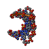 8f27C 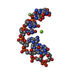 7kw4S S: Starting model for refinement C: citing same article ( |
|---|---|
| Similar structure data |
-Links
 PDBj
PDBj- Assembly
| Deposited unit | 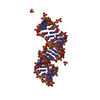
| ||||||||||||||||||||||||||||||
|---|---|---|---|---|---|---|---|---|---|---|---|---|---|---|---|---|---|---|---|---|---|---|---|---|---|---|---|---|---|---|---|
| 1 |
| ||||||||||||||||||||||||||||||
| Unit cell |
| ||||||||||||||||||||||||||||||
| Components on special symmetry positions |
|
-Components
| #1: RNA chain | Mass: 2540.588 Da / Num. of mol.: 6 / Source method: obtained synthetically / Source: (synth.) synthetic construct (others) #2: Chemical | ChemComp-SO4 /   Mass: 96.063 Da / Num. of mol.: 5 / Source method: obtained synthetically / Formula: SO4 Mass: 96.063 Da / Num. of mol.: 5 / Source method: obtained synthetically / Formula: SO4#3: Water | ChemComp-HOH / |  Mass: 18.015 Da / Num. of mol.: 19 / Source method: isolated from a natural source / Formula: H2O Mass: 18.015 Da / Num. of mol.: 19 / Source method: isolated from a natural source / Formula: H2OHas ligand of interest | Y | |
|---|
-Experimental details
-Experiment
| Experiment | Method: X-RAY DIFFRACTION / Number of used crystals: 1 |
|---|
- Sample preparation
| Crystal | Density Matthews: 2.66 Å3/Da / Density % sol: 53.73 % / Description: diamond shape |
|---|---|
| Crystal grow | Temperature: 293 K / Method: vapor diffusion, hanging drop Details: 2.0 M Ammonium sulfate, 0.1 M HEPES sodium pH 7.5, 2% v/v Polyethylene glycol 400 |
-Data collection
| Diffraction | Mean temperature: 99 K / Serial crystal experiment: N |
|---|---|
| Diffraction source | Source: SYNCHROTRON / Site: APS  / Beamline: 21-ID-F / Wavelength: 0.9787 Å / Beamline: 21-ID-F / Wavelength: 0.9787 Å |
| Detector | Type: RAYONIX MX-300 / Detector: CCD / Date: Oct 15, 2022 |
| Radiation | Protocol: SINGLE WAVELENGTH / Monochromatic (M) / Laue (L): M / Scattering type: x-ray |
| Radiation wavelength | Wavelength: 0.9787 Å / Relative weight: 1 |
| Reflection | Resolution: 2.27→30 Å / Num. obs: 2795 / % possible obs: 98.5 % / Redundancy: 8.2 % / CC1/2: 0.983 / CC star: 0.996 / Rmerge(I) obs: 0.074 / Rpim(I) all: 0.021 / Rrim(I) all: 0.077 / Rsym value: 0.068 / Χ2: 1.185 / Net I/σ(I): 30.1 |
| Reflection shell | Resolution: 2.27→2.31 Å / Redundancy: 8.4 % / Rmerge(I) obs: 0.874 / Mean I/σ(I) obs: 4.2 / Num. unique obs: 709 / CC1/2: 0.935 / CC star: 0.983 / Rpim(I) all: 0.224 / Rrim(I) all: 0.903 / Rsym value: 0.675 / Χ2: 1.088 / % possible all: 99.7 |
- Processing
| Software |
| ||||||||||||||||||||||||||||||||||||||||||||||||||||||||||||||||||||||||||||||||||||||||||||||||||||||||||||||||||||||||||||||||||||||||||||||||||||||||||||||||||||||||||||||||||||||
|---|---|---|---|---|---|---|---|---|---|---|---|---|---|---|---|---|---|---|---|---|---|---|---|---|---|---|---|---|---|---|---|---|---|---|---|---|---|---|---|---|---|---|---|---|---|---|---|---|---|---|---|---|---|---|---|---|---|---|---|---|---|---|---|---|---|---|---|---|---|---|---|---|---|---|---|---|---|---|---|---|---|---|---|---|---|---|---|---|---|---|---|---|---|---|---|---|---|---|---|---|---|---|---|---|---|---|---|---|---|---|---|---|---|---|---|---|---|---|---|---|---|---|---|---|---|---|---|---|---|---|---|---|---|---|---|---|---|---|---|---|---|---|---|---|---|---|---|---|---|---|---|---|---|---|---|---|---|---|---|---|---|---|---|---|---|---|---|---|---|---|---|---|---|---|---|---|---|---|---|---|---|---|---|
| Refinement | Method to determine structure: MOLECULAR REPLACEMENT Starting model: 7KW4 Resolution: 3.2→19.94 Å / Cor.coef. Fo:Fc: 0.923 / Cor.coef. Fo:Fc free: 0.835 / SU B: 20.47 / SU ML: 0.35 / Cross valid method: THROUGHOUT / ESU R Free: 0.598 / Stereochemistry target values: MAXIMUM LIKELIHOOD / Details: HYDROGENS HAVE BEEN ADDED IN THE RIDING POSITIONS
| ||||||||||||||||||||||||||||||||||||||||||||||||||||||||||||||||||||||||||||||||||||||||||||||||||||||||||||||||||||||||||||||||||||||||||||||||||||||||||||||||||||||||||||||||||||||
| Solvent computation | Ion probe radii: 0.8 Å / Shrinkage radii: 0.8 Å / VDW probe radii: 1.2 Å / Solvent model: MASK | ||||||||||||||||||||||||||||||||||||||||||||||||||||||||||||||||||||||||||||||||||||||||||||||||||||||||||||||||||||||||||||||||||||||||||||||||||||||||||||||||||||||||||||||||||||||
| Displacement parameters | Biso mean: 19.063 Å2
| ||||||||||||||||||||||||||||||||||||||||||||||||||||||||||||||||||||||||||||||||||||||||||||||||||||||||||||||||||||||||||||||||||||||||||||||||||||||||||||||||||||||||||||||||||||||
| Refinement step | Cycle: 1 / Resolution: 3.2→19.94 Å
| ||||||||||||||||||||||||||||||||||||||||||||||||||||||||||||||||||||||||||||||||||||||||||||||||||||||||||||||||||||||||||||||||||||||||||||||||||||||||||||||||||||||||||||||||||||||
| Refine LS restraints |
|